Features
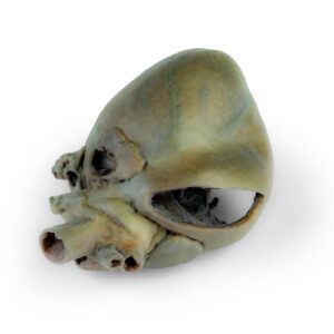
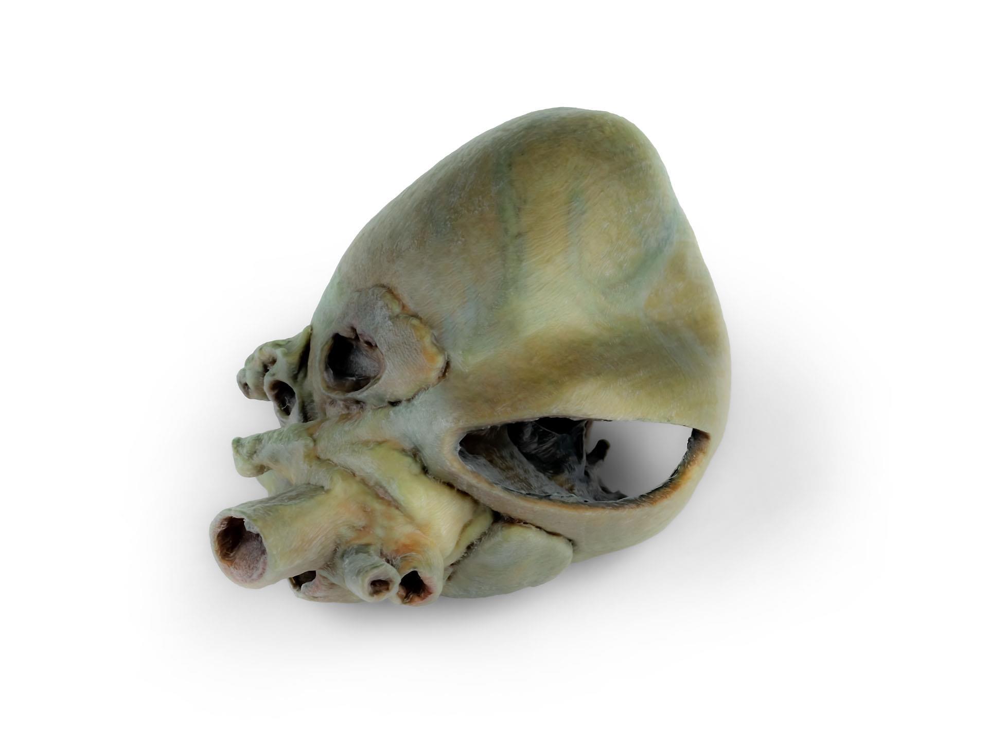
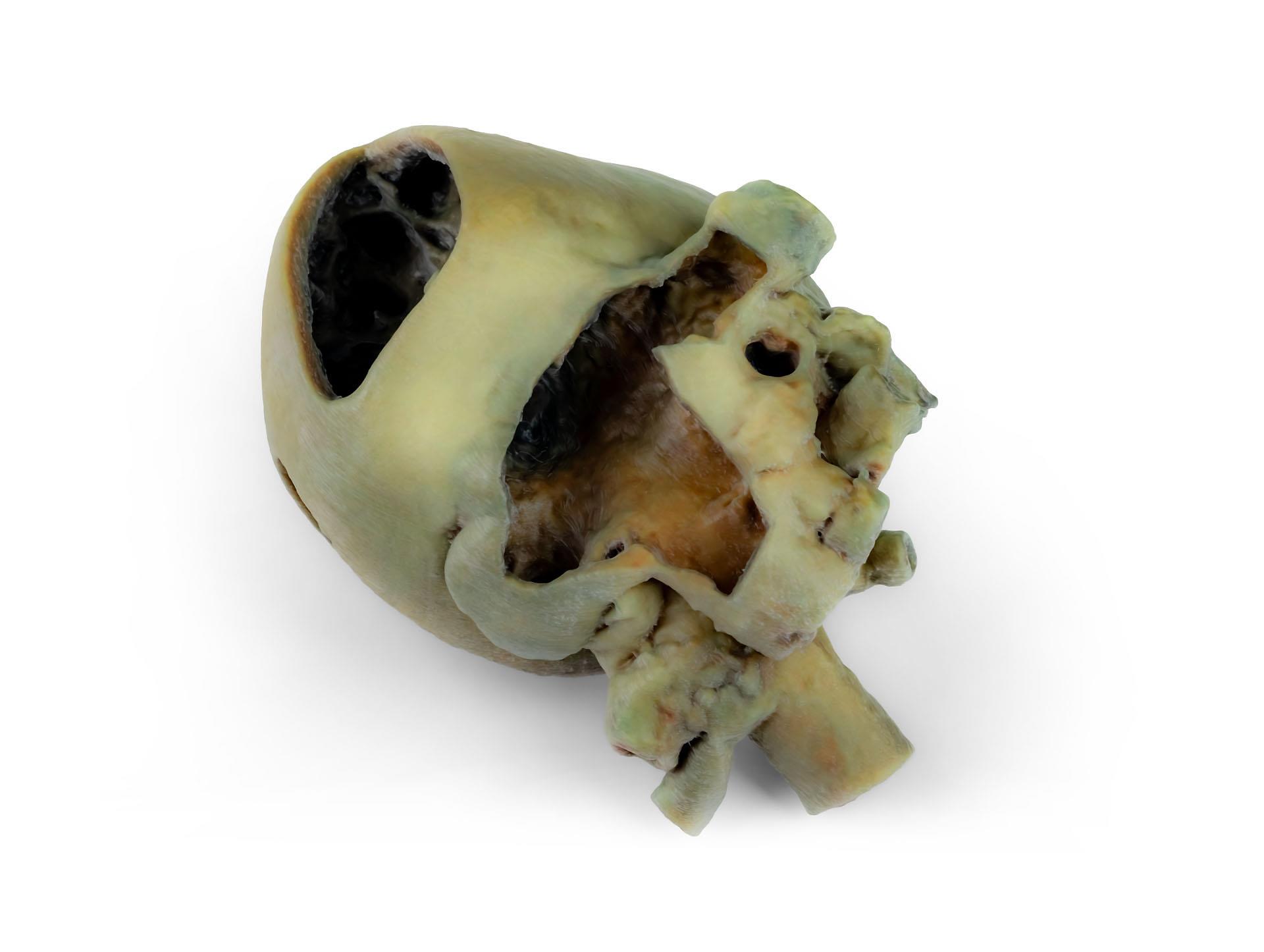
The Canine heart with dissection is a clinical-grade anatomical specimen designed for veterinary educators, students, and professionals. It offers an accurate and detailed look inside a canine heart, helping users learn, visualize, and teach cardiac anatomy with confidence. Choose this product to bring peerless clarity and realism to your veterinary studies or training sessions.
Explore Canine Cardiac Anatomy in Stunning Detail with the Canine heart with dissection
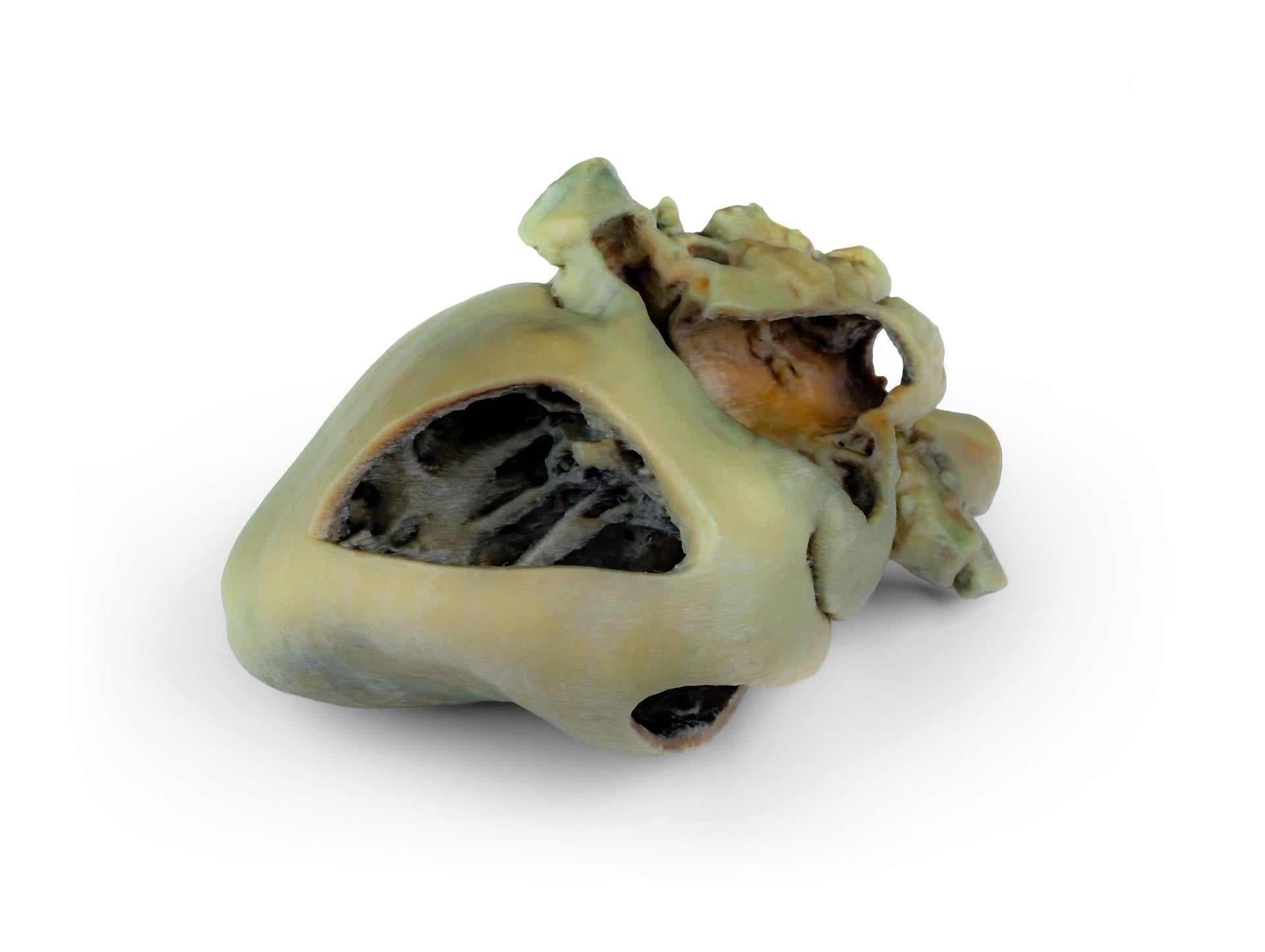
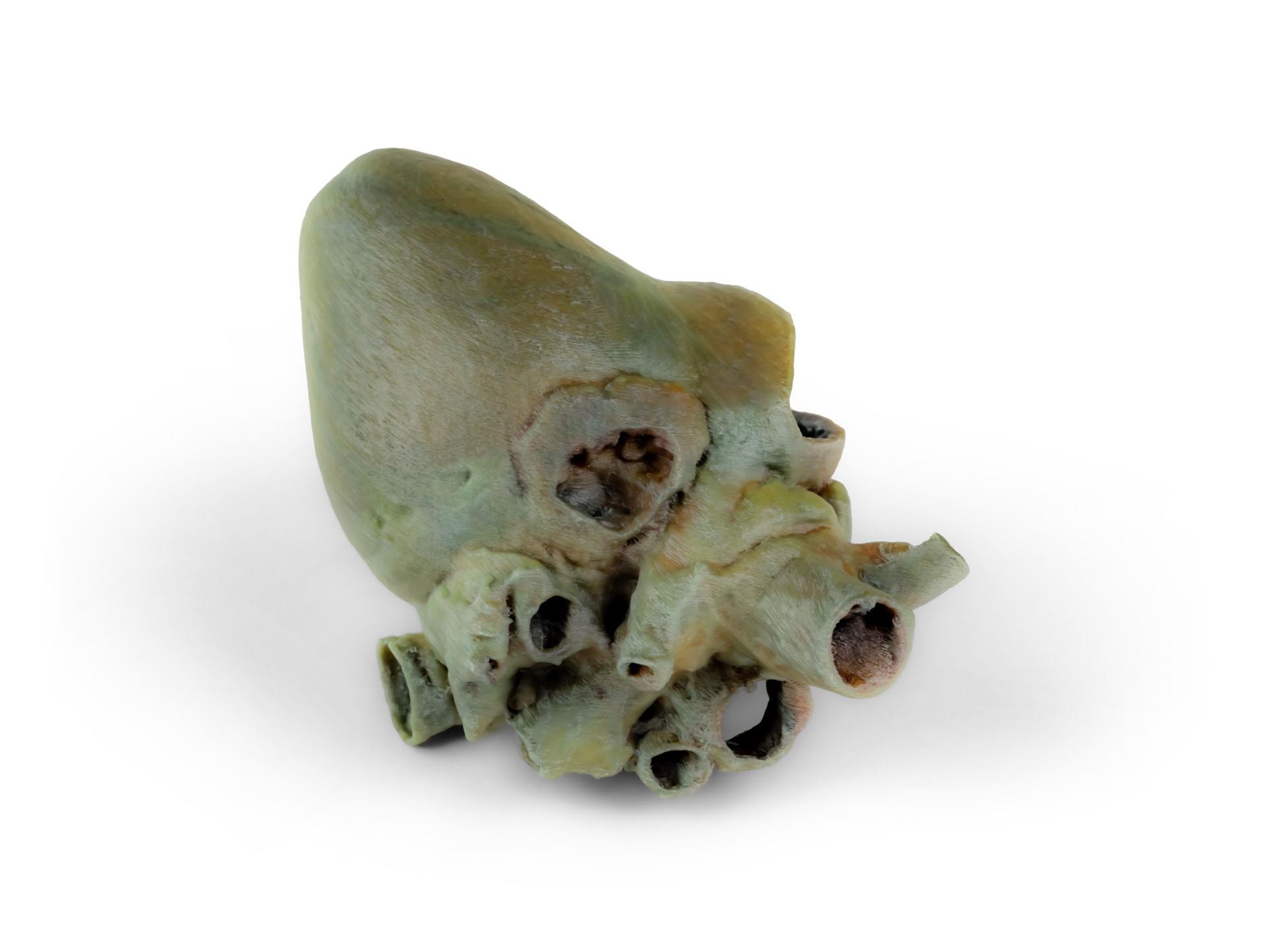
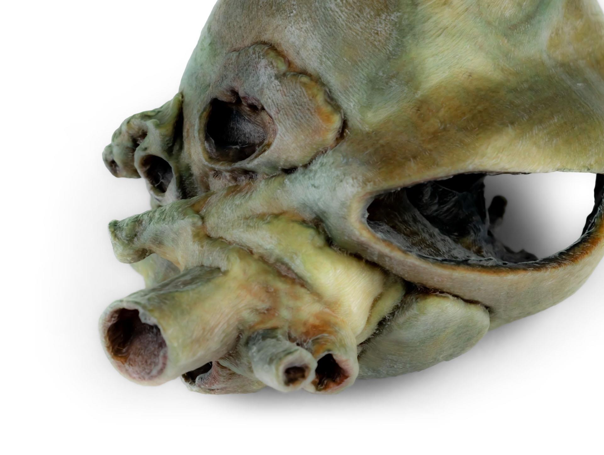
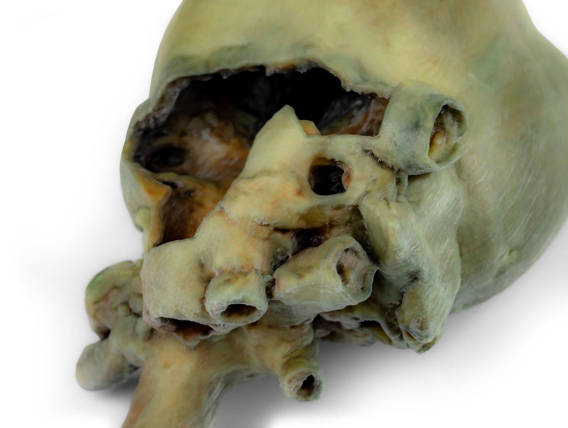
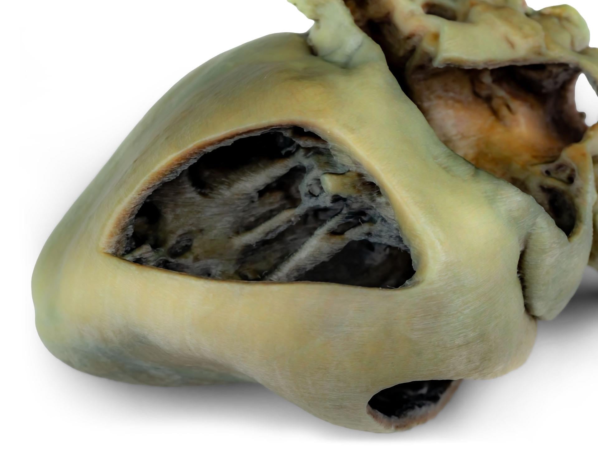
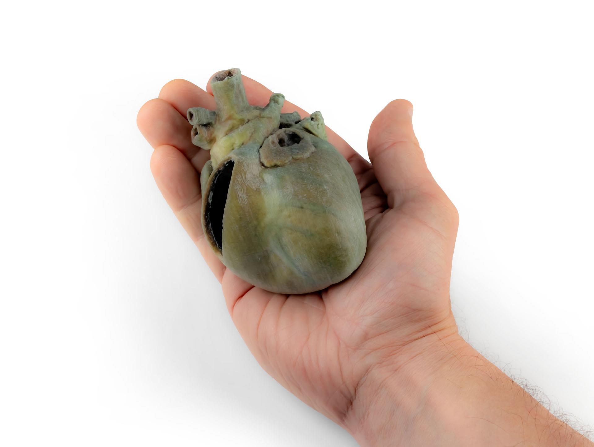
This expertly dissected specimen delivers exceptional insight into the internal and external features of the canine heart. The model shows both atrial and auricular faces, and reveals the base of the heart where the ascending aorta and pulmonary trunk connect. You can examine the entry points of the cranial and caudal venae cavae into the right atrium. The pulmonary trunk’s bifurcation and pulmonary veins entering the left atrium stand out clearly. Interventricular and coronary grooves are visible for added learning depth. Key internal structures are on display, such as the right atrioventricular (tricuspid) valve with chordae tendineae, papillary muscles, septomarginal trabecula, and open atrial walls, making this a comprehensive teaching resource.
Features and Benefits
- Displays full external heart anatomy including atrial and auricular faces
- Shows ascending aorta, pulmonary trunk and their relationship
- Clearly visible entry of cranial and caudal venae cavae into right atrium
- Pulmonary trunk bifurcation into pulmonary arteries
- Termination of pulmonary veins into left atrium is clear
- Exposes interventricular and coronary grooves
- Right atrial wall removed to reveal tricuspid valve, chordae tendineae and papillary muscles
- Features septomarginal trabecula (moderator band) inside right ventricle
- Open left atrial wall displays internal lumen
- Clinical-grade quality for educational reliability
Indications for Use
- Veterinary medical education and training
- Anatomy demonstration for students and professionals
- Hands-on learning for clinical or academic environments
Size Guide
- Single clinical specimen – typical adult canine heart size