Features
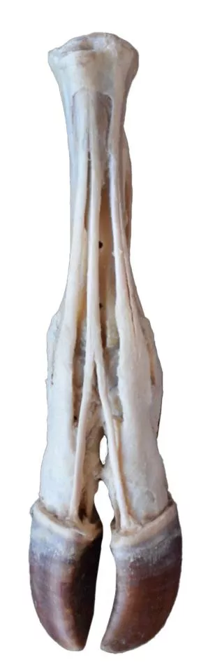
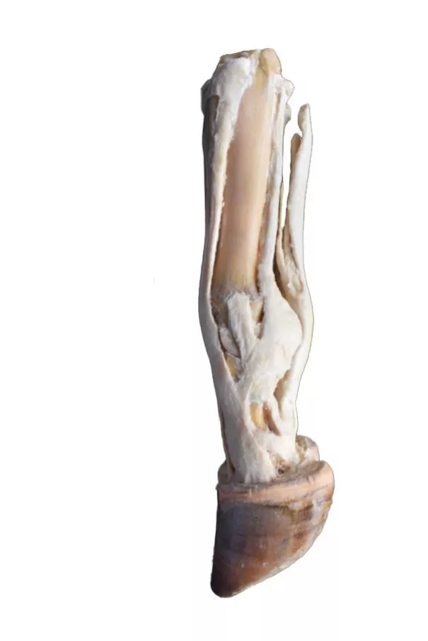
The Ox foot – tendons and ligaments is a clinical-quality anatomical specimen of a right ox foot designed for veterinary educators, students, and professionals. It provides a meticulous view of tendon and ligament structures, making it ideal for anatomical study and teaching. Choose this model to ensure accurate veterinary training with real anatomical detail that other models or illustrations cannot match.
Unlock Precision in Veterinary Learning with the Ox foot – tendons and ligaments
This premium specimen captures the full anatomy of an ox’s right foot, from the metatarsus down to the distal phalanges. On the dorsal side, you immediately see clear insertions of the common, lateral, and medial digital extensor tendons. The plantar aspect reveals detailed relationships of the superficial and deep digital flexor tendons, along with the interosseous ligament embedded in situ. Both capsular and extra-capsular ligaments for the metatarsophalangeal and interphalangeal joints remain intact, providing a comprehensive teaching tool. Veterinarians and educators can use this durable specimen for in-depth demonstrations and repeat handling, gaining unrivaled clarity into functional bovine limb anatomy.
Features and Benefits
- Displays a real ox’s right foot from metatarsus to distal phalanges
- Shows insertion of common, lateral, and medial digital extensor tendons on dorsal side
- Presents relationships of superficial and deep digital flexor tendons with interosseous ligament on plantar aspect
- Preserves both capsular and extra-capsular ligaments of metatarsophalangeal and interphalangeal joints
- Clinical-grade specimen crafted for vet education and detailed demonstration
Indications for Use
- Veterinary teaching and anatomical study
- Hands-on demonstration of bovine foot structures
- Training for professionals and students in large animal anatomy
Size Guide
- Full ox right foot specimen from metatarsus to distal phalanges
- Includes all tendons and ligaments as described