



Features
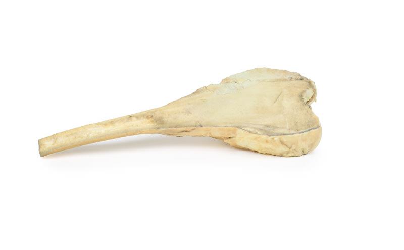
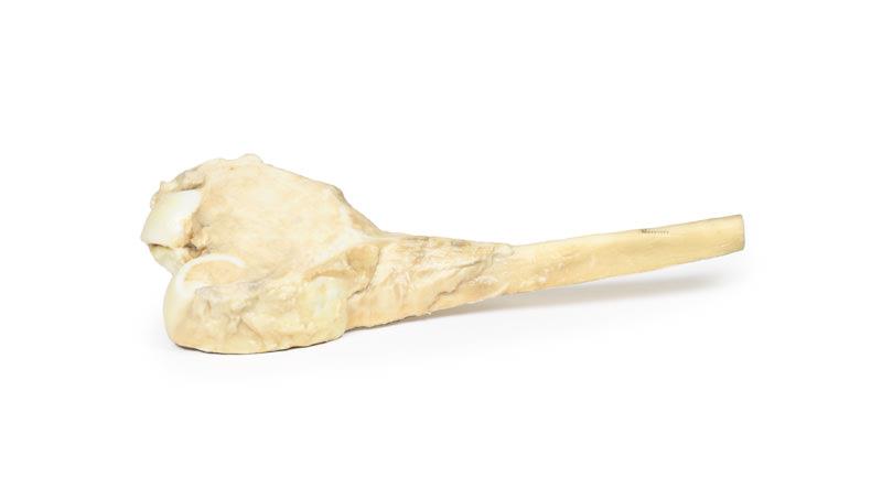
The Osteosarcoma of femur is a clinical-grade 3D printed anatomical model designed for medical professionals, educators, and students. It vividly demonstrates the pathological features of a femoral osteosarcoma, aiding in education and advanced research. Choose this product for precise, hands-on understanding of malignant bone tumors using accurate and high-quality anatomical replication.
Enhance Medical Education with Realistic Tumor Anatomy
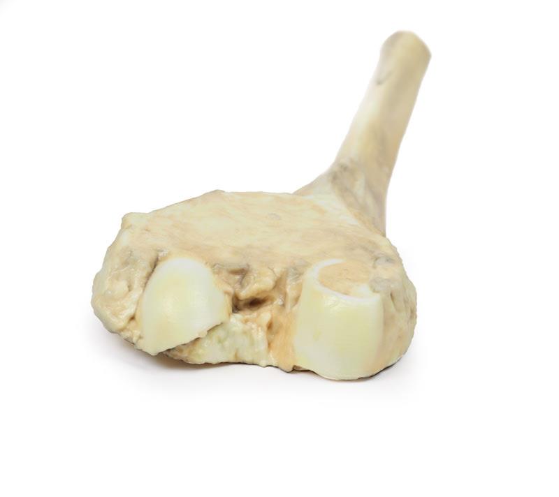
Gain a deeper understanding of primary malignant bone tumors with the Osteosarcoma of femur. This model replicates the distal femur excised from a clinical case involving a 16-year-old male patient. It highlights the pale, infiltrative tumor extending through the periosteum, demonstrating classic features including periosteal reaction and tumor spread near the articular surface. Incorporate this high-fidelity tool into medical teaching scenarios, enabling users to visualize, explore, and discuss osteosarcoma characteristics and management in detail.
Features and Benefits
- Life-sized 3D printed model based on real clinical pathology
- Displays large infiltrating tumor with periosteal extension (10 cm diameter)
- Shows periosteal reactive changes and tumor spread near the articular surface
- Educational focus on primary bone malignancy of the distal femur
- Robust, durable materials suitable for repeated handling in educational settings
- Clinical-grade detail for accurate pathology demonstration
Indications for Use
- Medical and health science education
- Tumor board and pathology teaching
- Surgical planning and anatomical demonstrations
- Research into primary bone tumors and tumor progression
Size Guide
- Full distal femur anatomical model
- Tumor size approx. 10 cm in greatest diameter as per clinical specimen
- Life-sized, representing actual human femoral anatomy

