Features
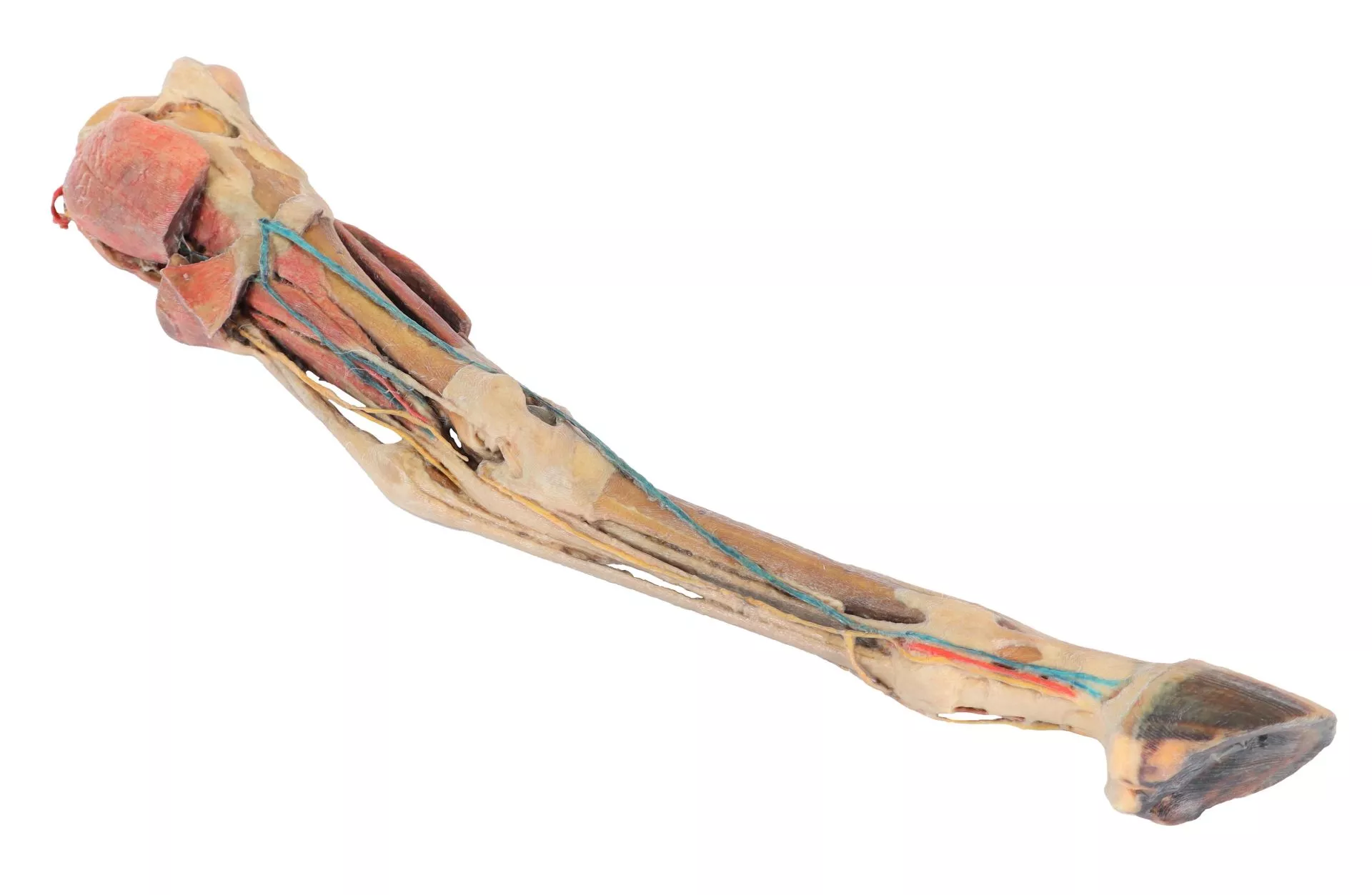
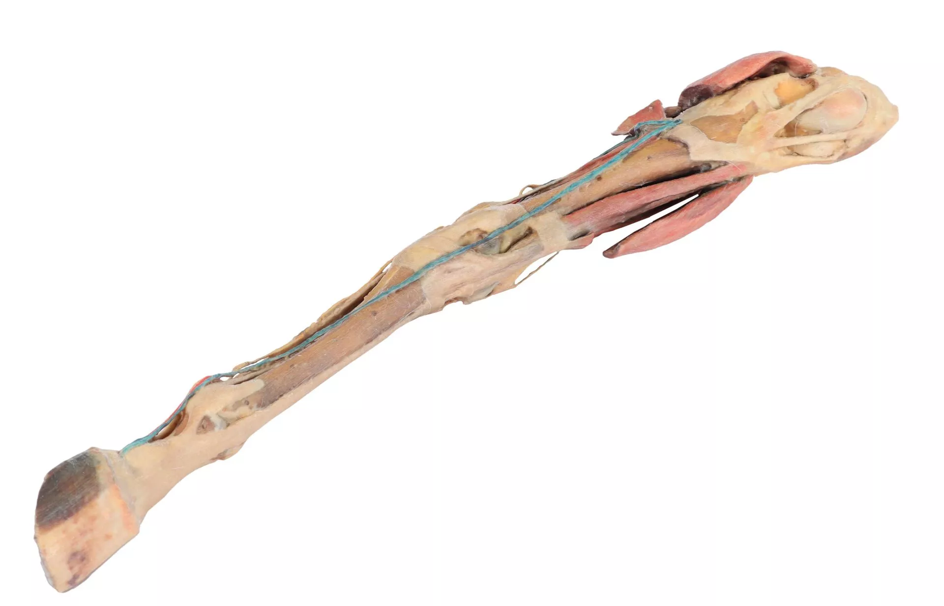
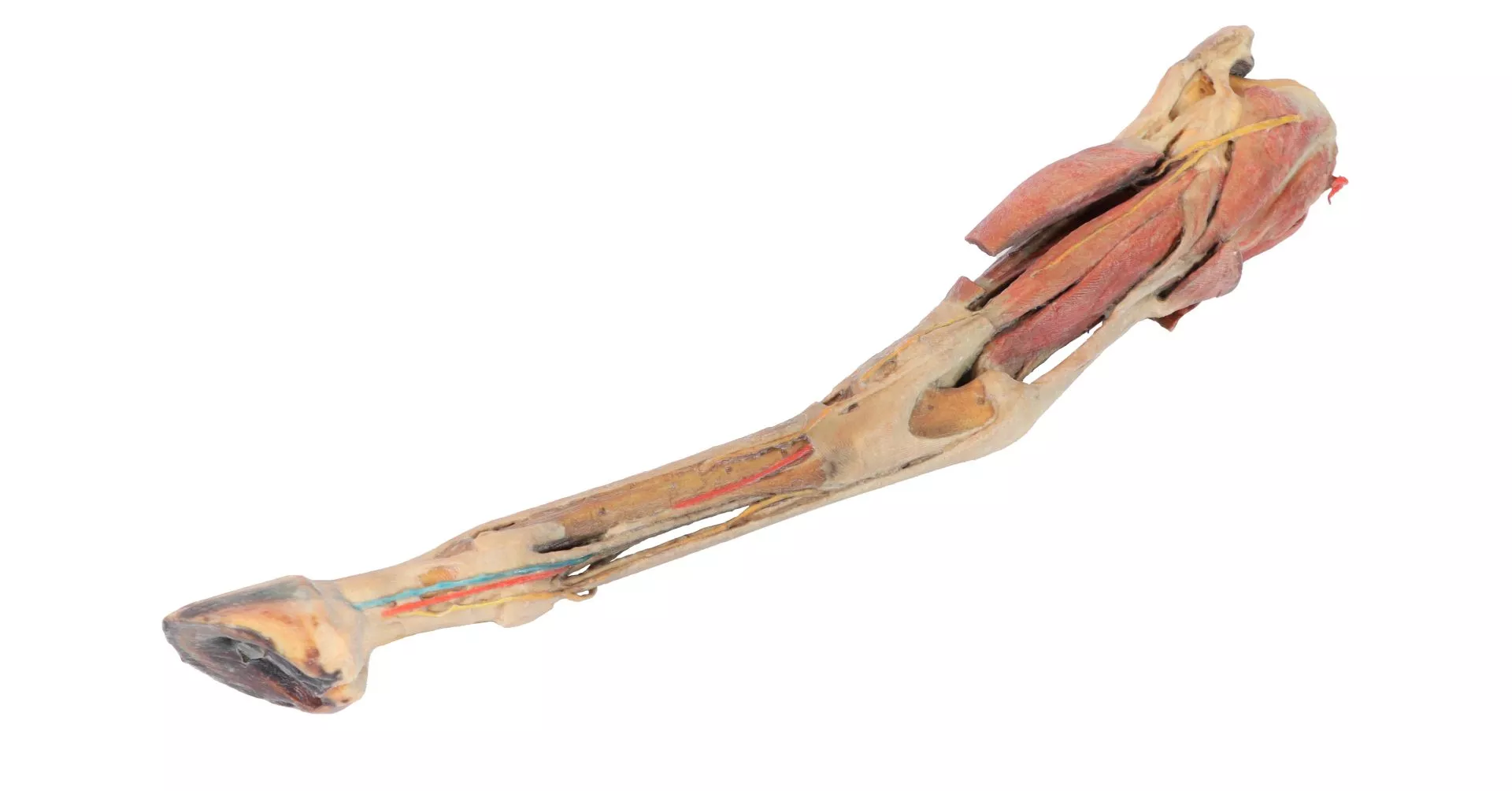
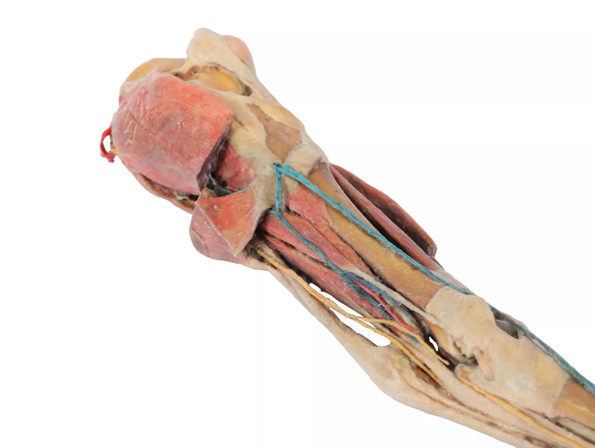
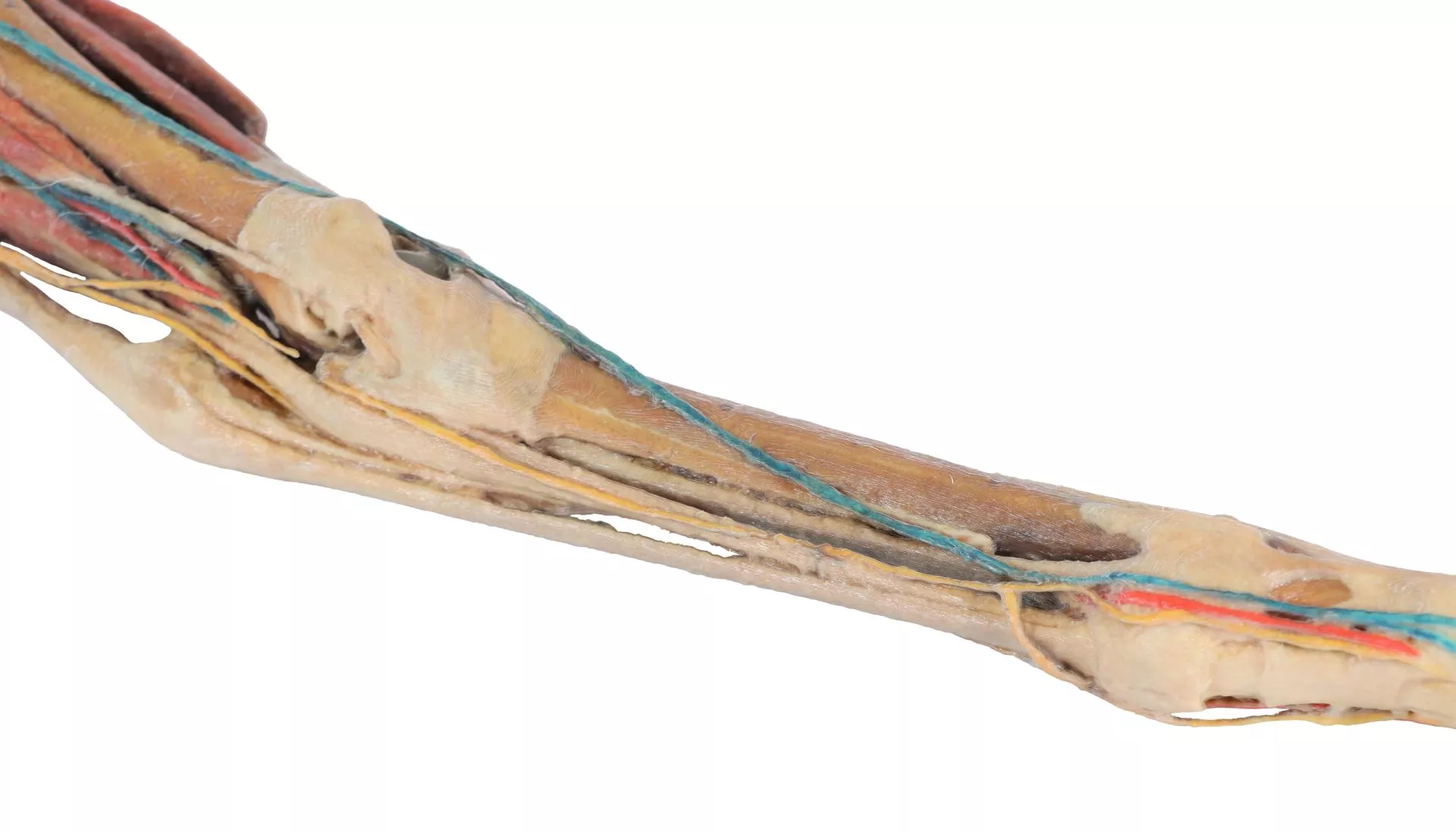
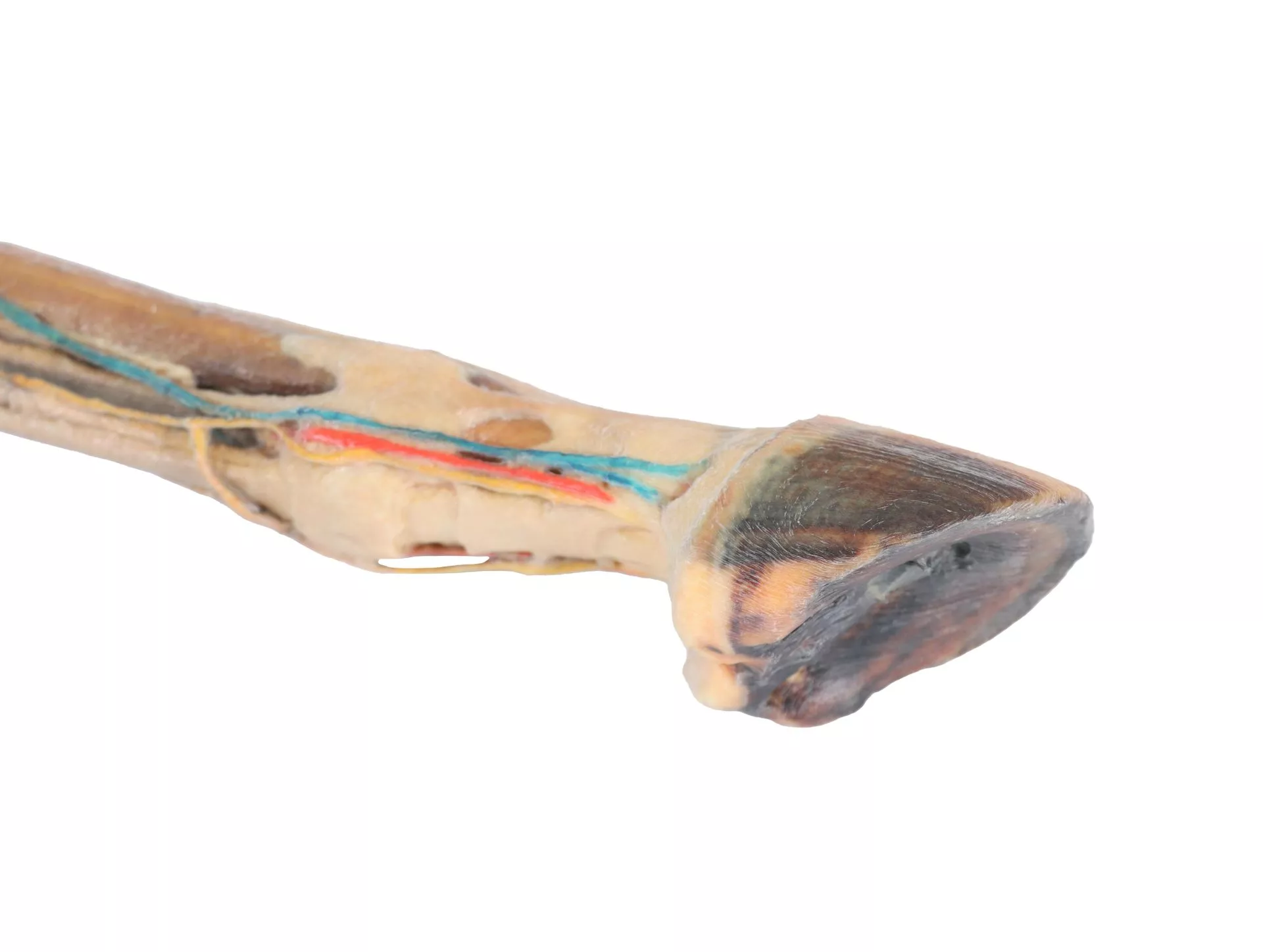
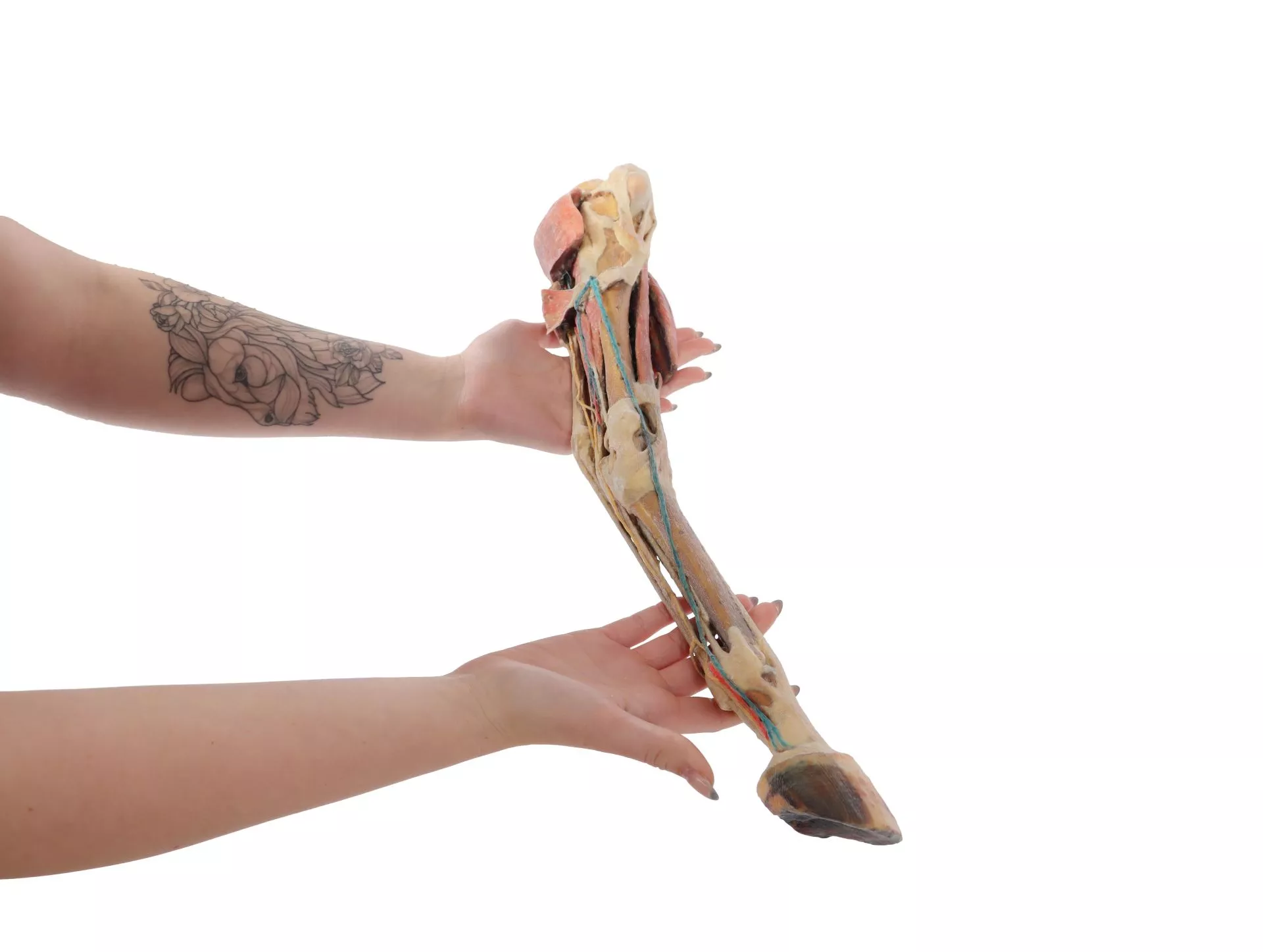
The Horse hindlimb – muscles, tendons, ligaments, vessels and nerves distal to the stifle – 1/3 life-size is a clinically detailed anatomical specimen for veterinary students and professionals. It simplifies understanding complex equine lower limb anatomy, enhancing study and surgical training. Choose this model for uncompromised precision and realistic features that elevate anatomical learning.
Unlock True-to-Life Equine Limb Anatomy for Veterinary Education
This horse hindlimb specimen displays muscles, tendons, ligaments, vessels, and nerves from the midthigh to the hoof at a third of their real size. Trainers and students can explore meticulously dissected stifle and tarsus joints, revealing menisci and essential ligaments. You see nerve and vessel pathways expertly presented in the popliteal fossa, tarsus, and digits. Use the Horse hindlimb – muscles, tendons, ligaments, vessels and nerves distal to the stifle – 1/3 life-size for hands-on learning and precise anatomical reference in lectures, clinics, and specialized equine courses.
Features and Benefits
- Superficial dissection of the hindlimb musculature from midthigh to hoof
- Exposed stifle and tarsus joint capsules showing menisci and main ligaments
- Extensor and flexor muscles revealed from femur and tibia origins to insertions in tarsus, metatarsus, and phalanges
- Nerves and vessels dissected in popliteal fossa, tarsus, and digit regions
- 1/3 life-size for easy handling and display
- Clinical-grade preparation for educational and professional settings
- Detailed descriptions available for advanced study
Indications for Use
- Veterinary anatomical education
- Surgical training and planning
- Classroom demonstration and lectures
- Professional anatomical reference
Size Guide
- 1/3 life-size specimen from midthigh to hoof
- Accurately scaled to one-third horse limb measurements for optimal demonstration