
Features
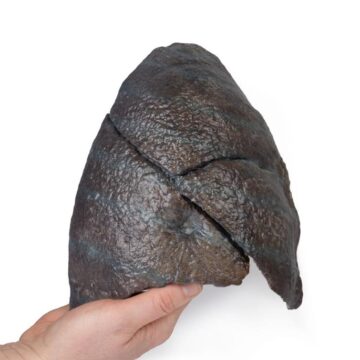
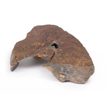
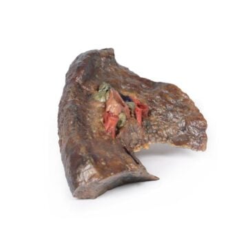
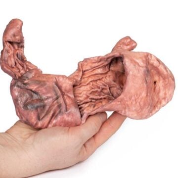
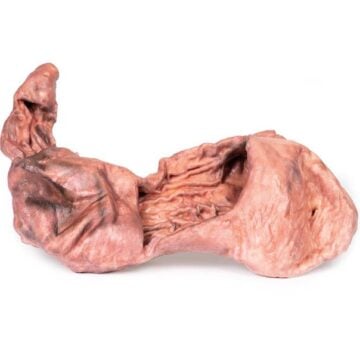
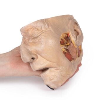
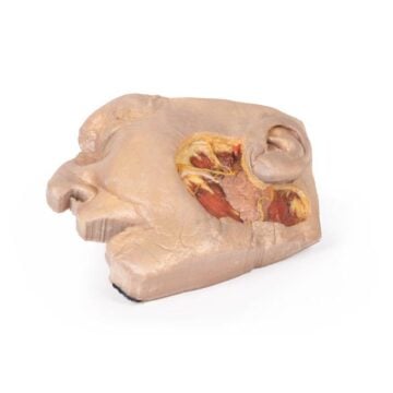
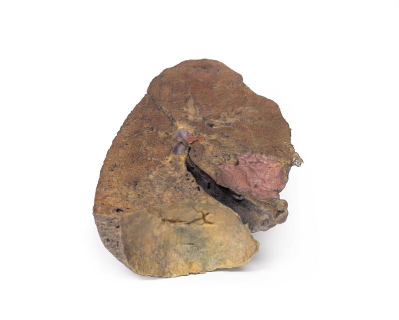
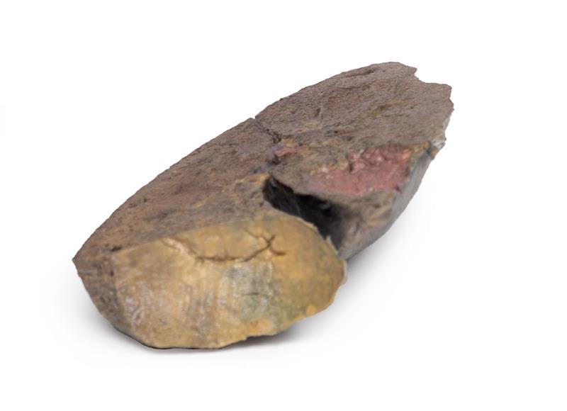
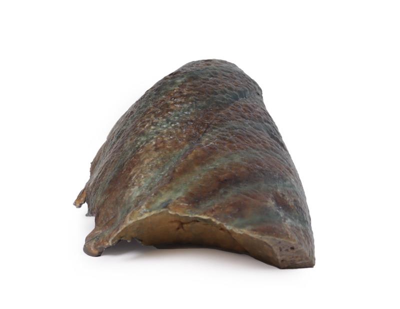
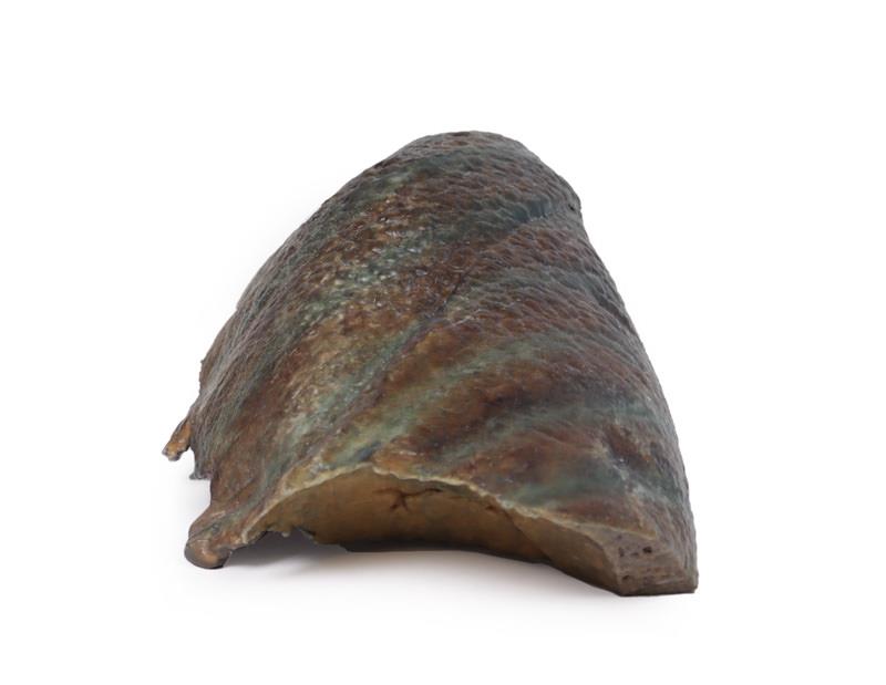
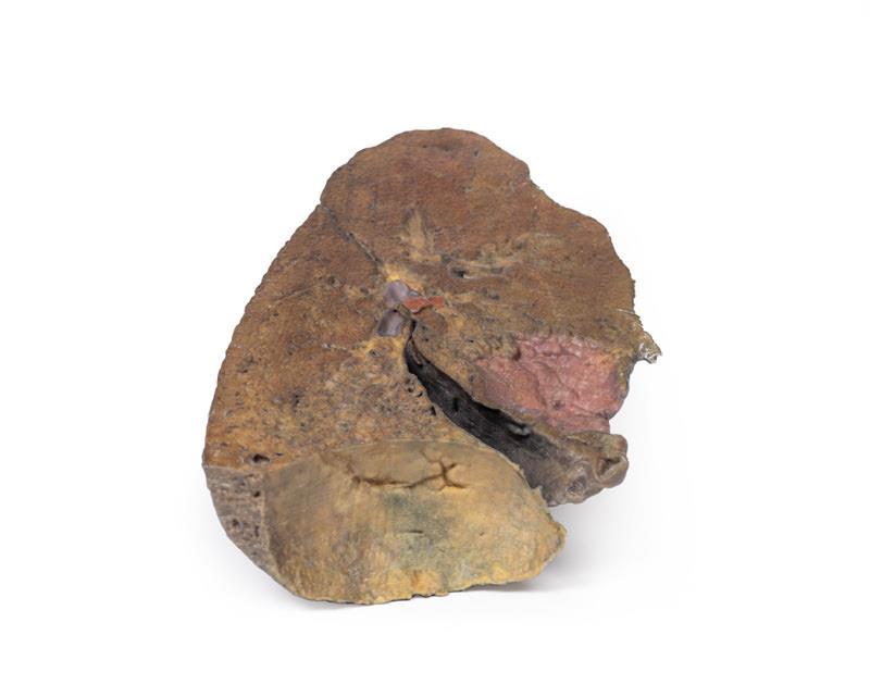
The Lung Slab, Hilum removed is a 3D printed anatomical model designed for medical educators, students, and healthcare professionals. It offers a unique view of pulmonary anatomy by presenting a dissected lung slab where the hilum has been removed. Choose this model for an unprecedented look at internal lung structures and better understanding of anatomical relationships.
Enhance Anatomy Lessons with Detailed 3D Pulmonary Visualization
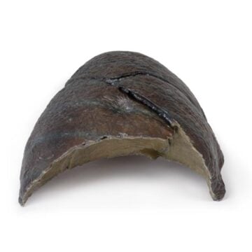
The Lung Slab, Hilum removed showcases intricate pulmonary details that improve the clarity of classroom demonstrations. By dissecting the lung along a parasagittal plane, this model reveals vascular and bronchial entry points while omitting the mediastinal surface. The cardiac impression stands out as the most concave region, clearly formed by the left ventricle. This model allows users to examine lung orientation above the diaphragm and understand the formation of the diaphragmatic surface. Clinical-grade 3D printing ensures lifelike accuracy and durability for repeated educational use, making it a top choice for institutions and anatomy instructors.
Features and Benefits
- 3D printed for clinical-grade accuracy and realism
- Parasagittal dissection removes mediastinal surface to reveal key internal structures
- Clear visualization of pulmonary arteries, veins, and bronchi at entry sites
- Prominent cardiac impression created by left ventricle
- Displays the lung’s diaphragmatic concavity
- Durable construction supports repeated educational use
- Ideal for teaching, demonstration, and anatomical study
Indications for Use
- Anatomy education in medical and allied health programs
- Classroom and laboratory demonstration of pulmonary and cardiac structures
- Preparation and review for surgical and clinical training
- Reference for pulmonary pathology discussions
Size Guide
- Single model – Actual anatomical size (adult lung specimen)
- Dimensions reflect real adult human lung, with parasagittal dissection and hilum not present