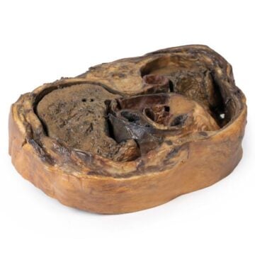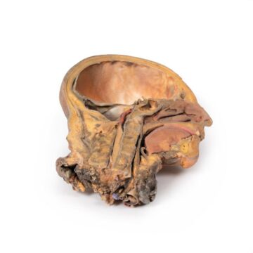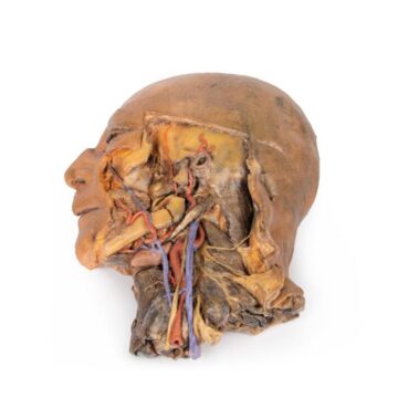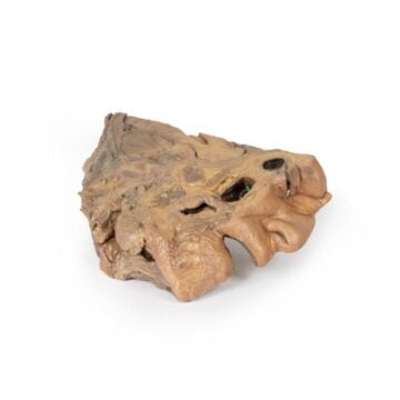
Features
The Brain (Cerebrum) is a highly detailed 3D printed anatomical model designed for medical educators, students, and professionals. It provides a hands-on solution for studying cerebral and meningeal organization without the complexities of cadaveric specimens. Choose this model for unmatched anatomical fidelity, reproducibility, and enhanced visual clarity in neuroanatomy education.
Discover Unmatched Anatomical Clarity with the Brain (Cerebrum)
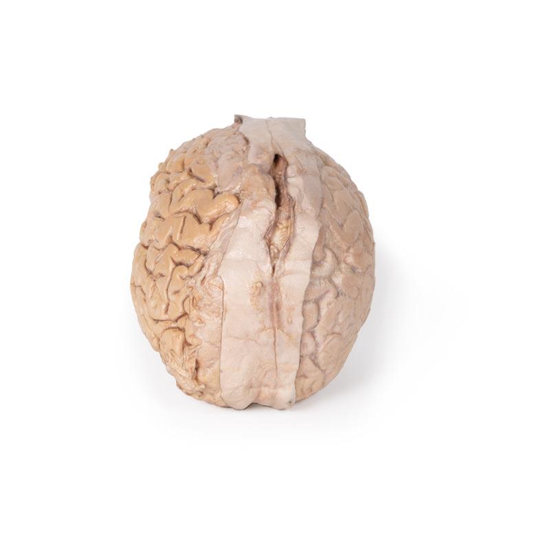
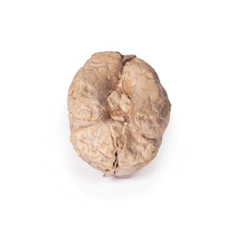
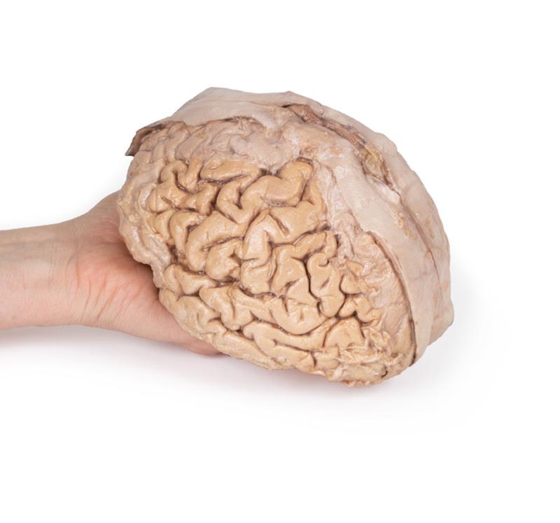
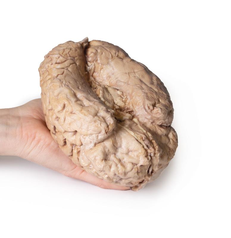
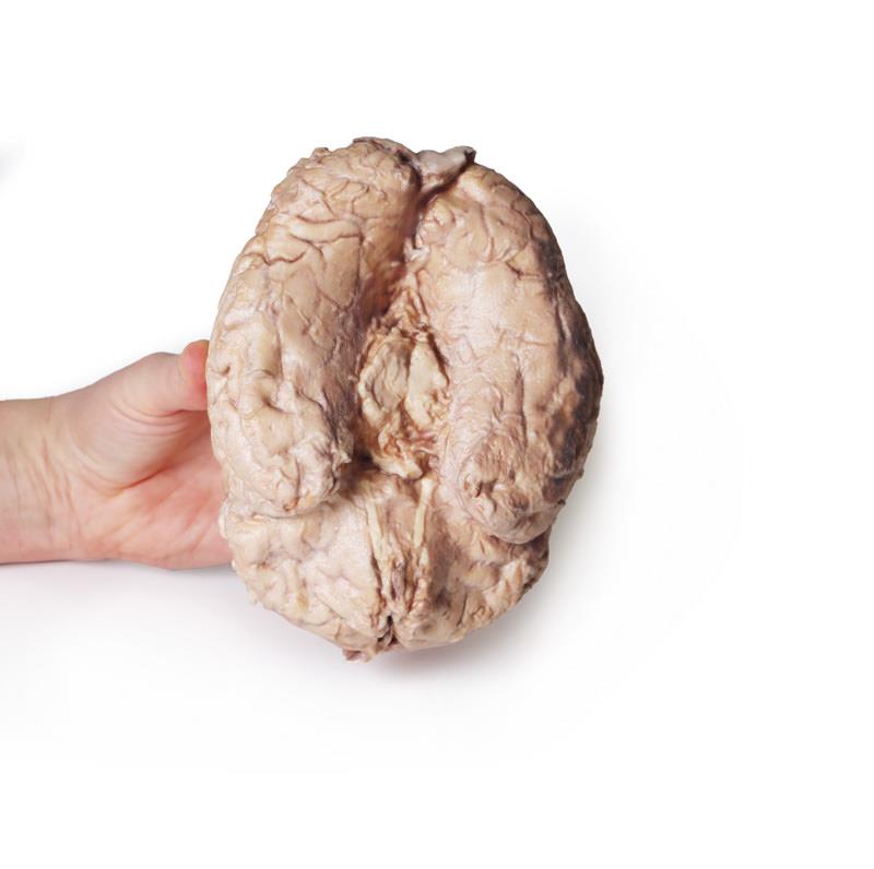
This Brain (Cerebrum) model stands out with its precise dissection between the left and right hemispheres, showcasing the relationship between brain structures and meninges as they appear in the cranial cavity. The model separates the cerebrum from the brainstem and cerebellum, features visible midbrain and cerebral peduncles on the inferior surface, and highlights olfactory tracts and bulbs. Varying dissection preserves the dura mater midline, exposes the superior sagittal sinus with clear visualization of arachnoid granulations, and demonstrates contrasting exposures of arachnoid and pia mater. These design elements give users direct views of the lateral and central sulci, essential for understanding cortical region boundaries. 3D-printed from real human anatomy, it offers safer handling, lower cost, and customization options compared to traditional specimens.
Features and Benefits
- 3D printed from real human anatomy for exceptional accuracy
- Clear separation of cerebrum from brainstem and cerebellum
- Visible parts of midbrain and cerebral peduncles on inferior surface
- Olfactory tracts and bulbs displayed on frontal lobe margin
- Differentiated dissection between cerebral hemispheres
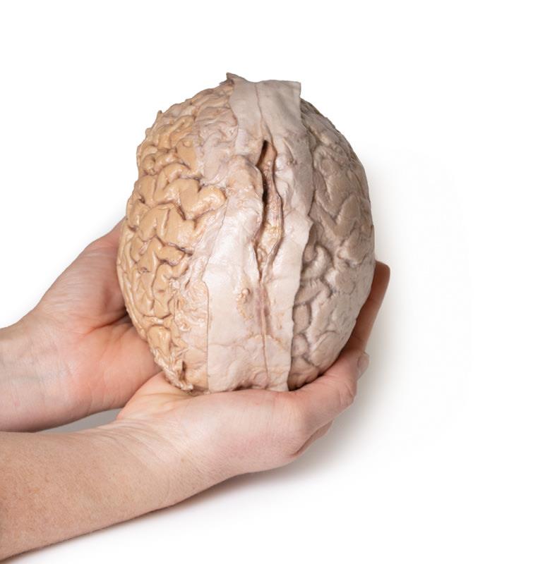
- Dura mater preserved and opened to show superior sagittal sinus
- Arachnoid granulations clearly visible
- Right hemisphere displays exposed arachnoid mater and vessels
- Left hemisphere exposes gyri and sulci covered with pia mater
- Distinct views of lateral and central sulci with defined lobe boundaries
- Consistent, reproducible standard for teaching
- Customizable to highlight specific structures or create cutaway views
- Ethically sound and easy to maintain compared to cadaveric specimens
Indications for Use
- Anatomy and neuroanatomy education in medical institutions
- Surgical and clinical reference for professionals
- Demonstration in lectures or practical training sessions
- Patient education or consultation support
- Curriculum standardization across teaching sites
Size Guide
- No sizing required — model depicts a full-scale adult human cerebrum with all features as described above.