Features
The Ventriculitis, Secondary to Septicaemia is a clinical-grade 3D printed anatomical model designed for medical educators, clinicians, and students. It demonstrates the pathology of ventriculitis as a complication of septicemia, providing exceptional detail for teaching, research, and study. Choose this model for its accuracy in representing critical neuropathological changes, enhancing your understanding of complex infectious diseases.
Elevate Neuroanatomy Teaching with the Ventriculitis, Secondary to Septicaemia Model
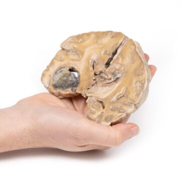
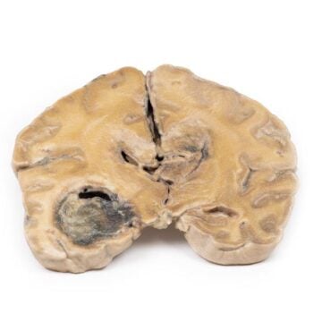
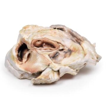
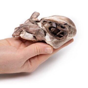
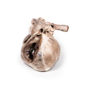
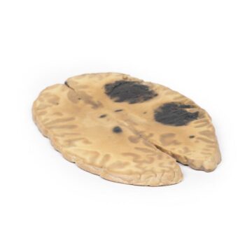
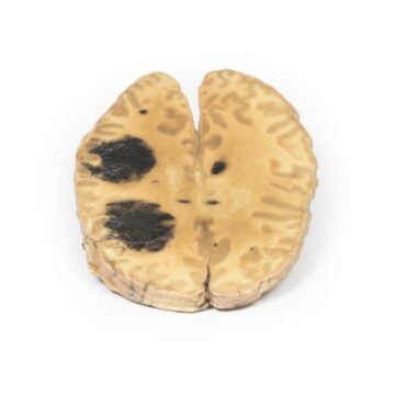
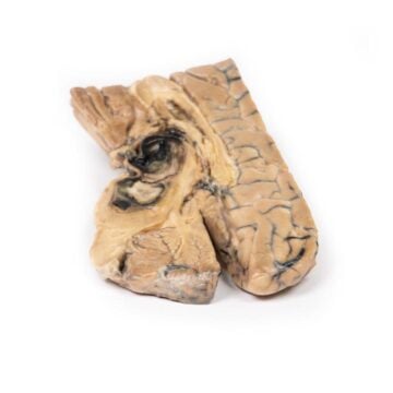
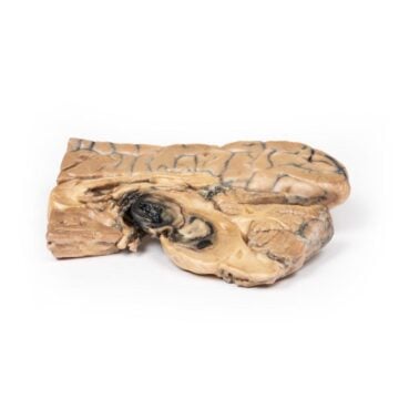
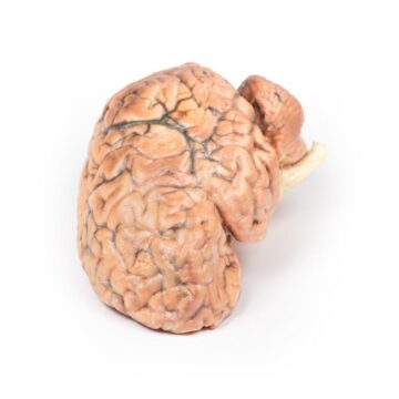
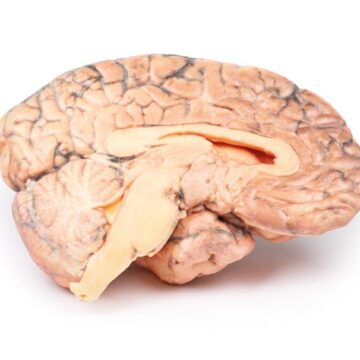
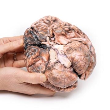
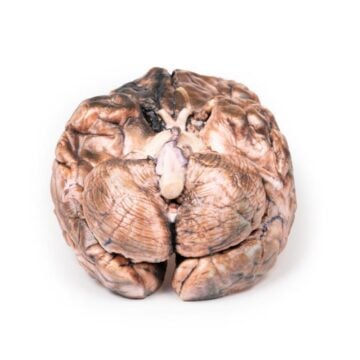
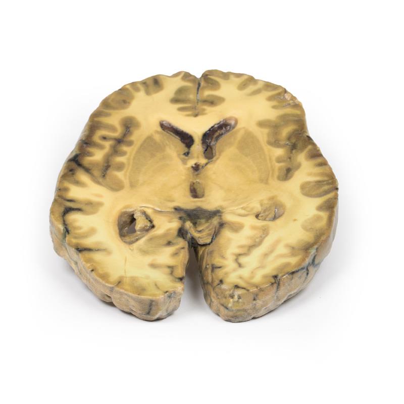
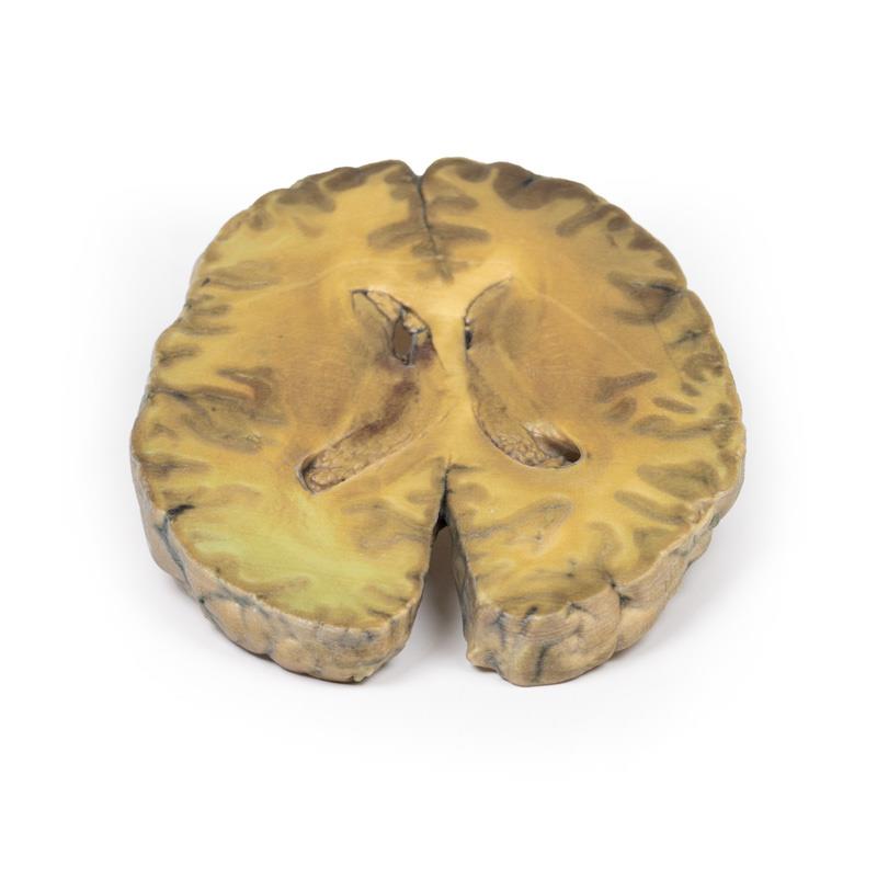
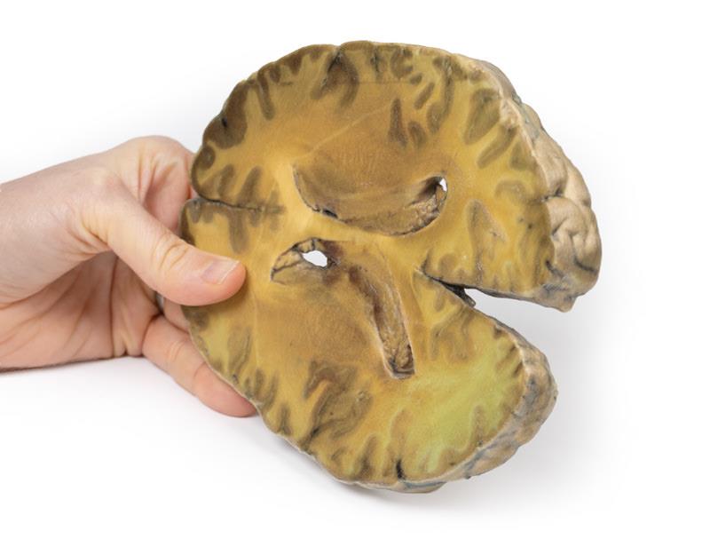
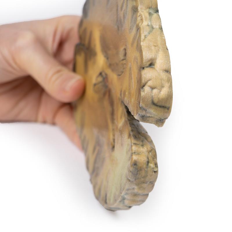
This meticulously crafted 3D printed model visualizes real-case ventriculitis, secondary to septicemia, as seen in the brain of a 50-year-old patient. It displays the gross changes found in both lateral ventricles, including a thickened, roughened ependymal lining and accumulations of cellular debris near the choroid plexus and anterior horn. The lower brain surface features and normal anatomy of the caudate nucleus, lentiform nucleus, and internal capsule stand out, making this an ideal tool for instruction. Detailed histological findings, such as extensive neutrophil infiltration, vascular involvement, and inflammation extending into cerebral parenchyma, reinforce the clinical value of this model. Educators and institutions benefit from a durable, clinical-grade, and highly accurate representation of rare neuropathology.
Features and Benefits
- 3D printed from clinical specimen for unparalleled anatomical detail
- Displays both lateral ventricles with realistic thickened ependymal lining
- Shows accurate debris accumulation at choroid plexus and anterior horn
- Reveals the arrangement of major brain structures including caudate and lentiform nuclei
- Highlights histological features such as neutrophil infiltration and vascular changes
- Clinical-grade, education standard construction designed for repeated handling
- Supports advanced neuropathology teaching and demonstration
Indications for Use
- Medical and health sciences education
- Pathology and neuroanatomy teaching
- Clinician and resident training on central nervous system infection complications
- Demonstrations of ventriculitis secondary to infection or medical intervention
- Reference for laboratory, imaging, and autopsy findings
Size Guide
- Model represents a life-size horizontal brain slice through both cerebral hemispheres
- Demonstrates full structures of both lateral ventricles, caudate nucleus, lentiform nucleus, and internal capsule as seen in adults
- SKU – ERLMP2008