

Features
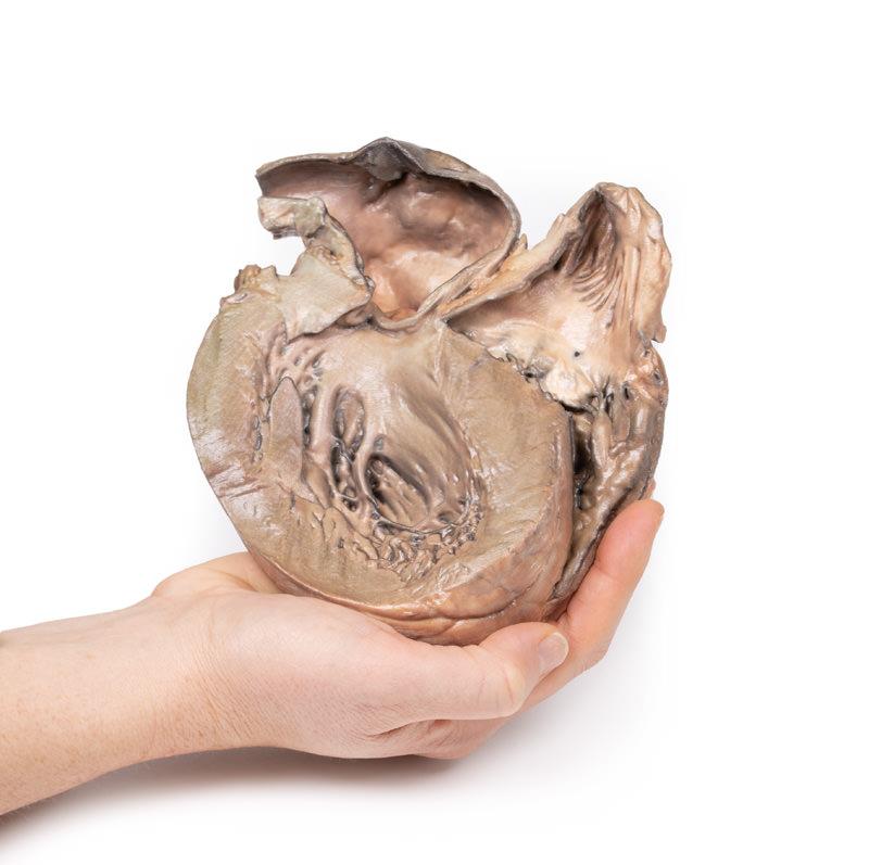
The Ruptured Thoracic Aortic Aneurysm is a 3D printed anatomical model designed for medical educators, students, and healthcare professionals. It provides a true-to-life representation of a saccular dilatation with rupture, supporting detailed visualization and hands-on learning. Choose this model for unparalleled accuracy when teaching complex cardiac and vascular anatomy.
Bring Cardiac Pathology to Life with a 3D Ruptured Thoracic Aortic Aneurysm Model
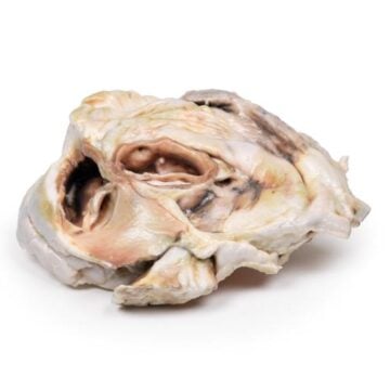
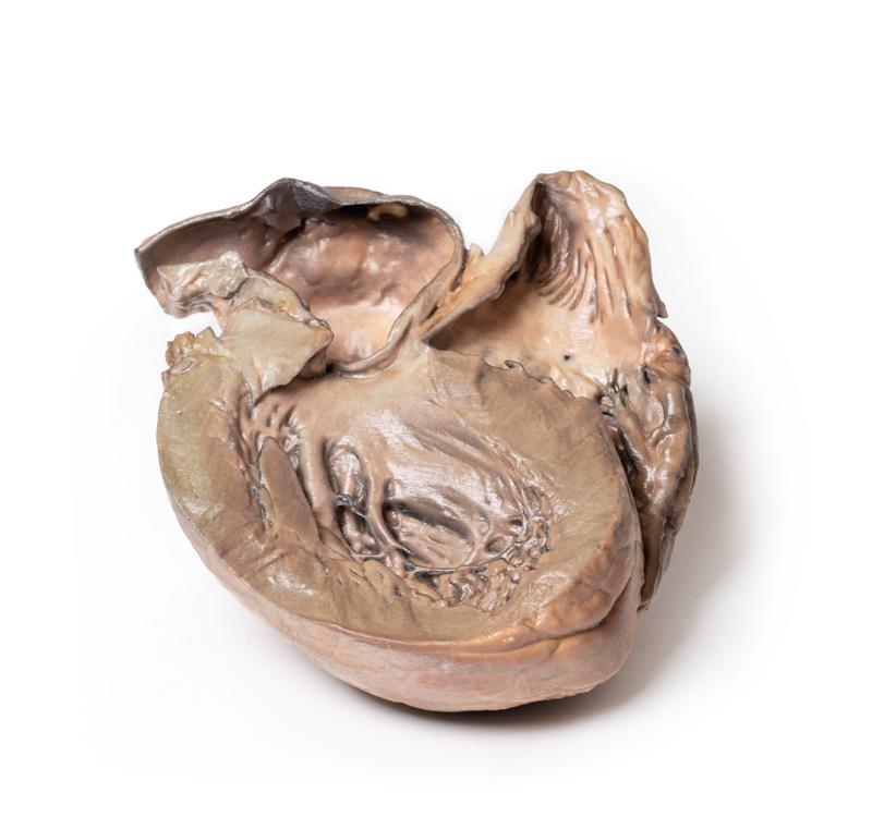
This clinical-grade Ruptured Thoracic Aortic Aneurysm model delivers exceptional anatomical detail for comprehensive study and demonstration. Expertly crafted in Australia using advanced 3D printing, the model captures the prominent saccular aneurysm in the thoracic ascending aorta, including surface atherosclerotic plaques and clear rupture markings. Both hypertrophied ventricles are displayed from the posterior aspect, while the coronary arteries and aortic and tricuspid valves remain anatomically normal. This durable teaching aid enables hands-on training for critical cardiac and vascular scenarios, making it indispensable for academic institutions and specialist settings.
Features and Benefits
- High-resolution 3D printed replica of a true ruptured thoracic aortic aneurysm
- Accurately displays hypertrophied ventricles seen from posterior
- Clear presentation of saccular dilatation with defined atherosclerotic plaques and rupture
- Normal coronary arteries and intact aortic and tricuspid valves for contrast
- Clinical-grade quality suitable for education and examination
- Produced in Australia with fast national delivery
- Robust construction for repeated handling in teaching scenarios
Indications for Use
- Medical education and training in cardiology and vascular pathology
- Demonstrating ascending aorta disease progression and rupture
- Simulation-based learning for healthcare professionals
- Patient education regarding aortic aneurysm conditions
Size Guide
- Full-sized anatomical 3D model representing entire heart with thoracic ascending aorta
- Actual dimensions and weight may vary as each print replicates clinical specimen scale