
Features
The Female right pelvis is a 3D printed anatomical model designed for educators, clinicians, and students who require detailed pelvic structure visualization. It provides accurate anatomical representation for teaching, study, or clinical demonstration. Choosing this model ensures access to premium educational tools with lifelike and precise dissection detail.
Unlock Advanced Anatomical Clarity with the Female right pelvis Model
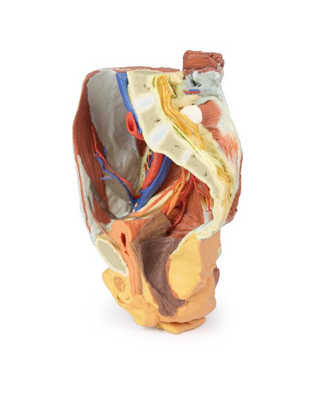
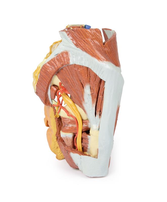
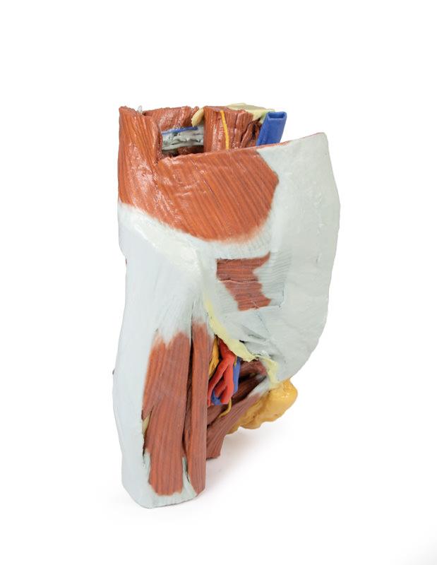
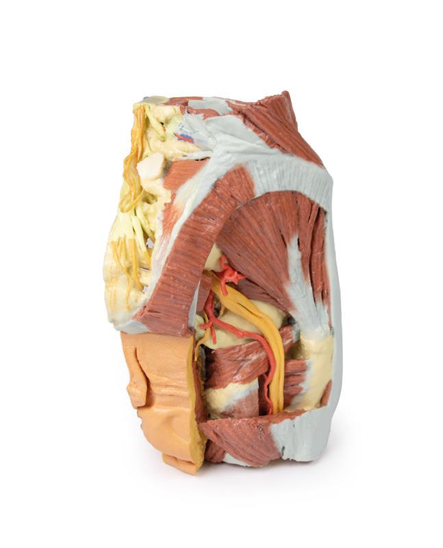
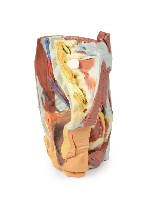
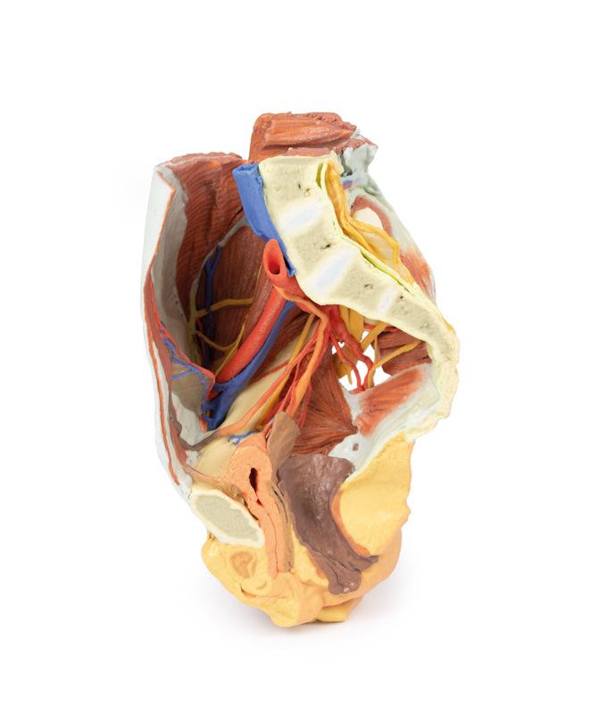
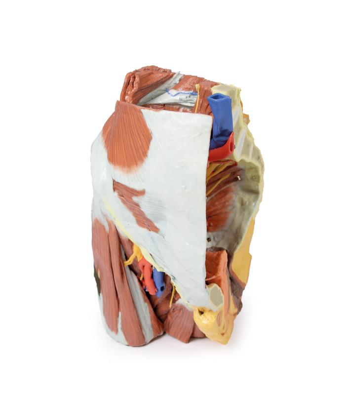
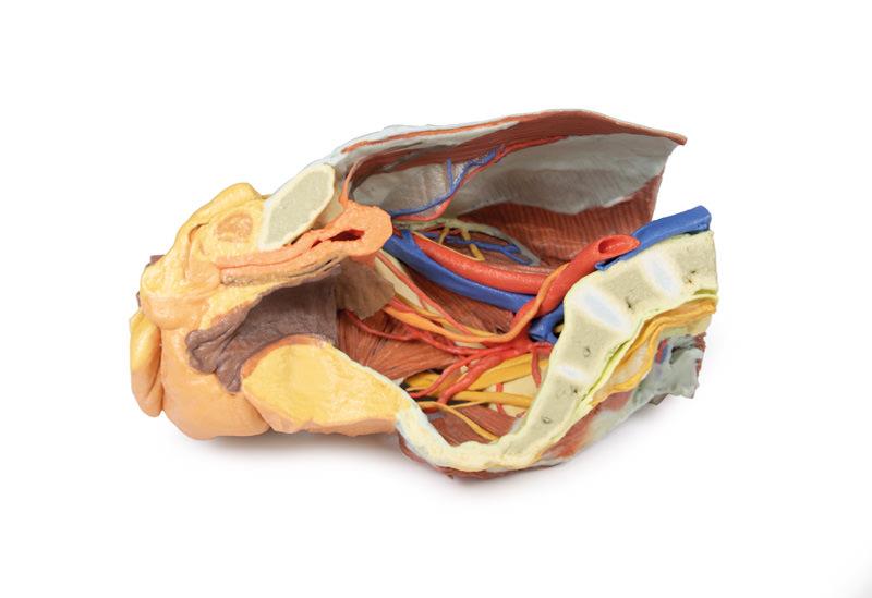
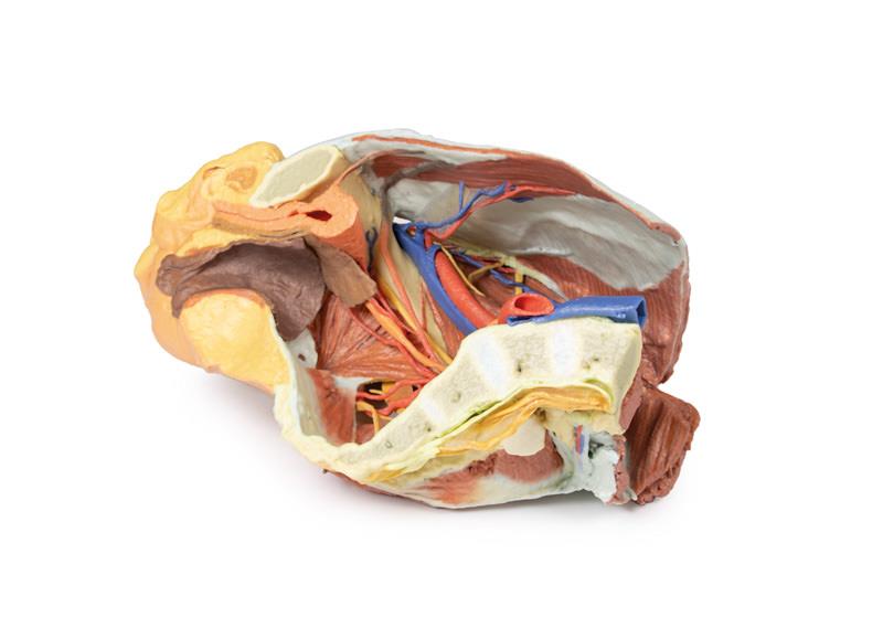
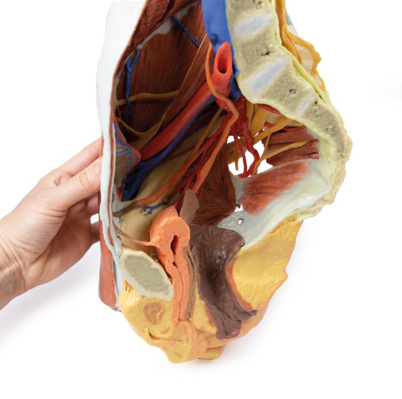
This clinical-grade 3D printed specimen offers unmatched insight into the female pelvic anatomy. Meticulously crafted, it displays the right half of the pelvis sectioned midsagittally and transversely at the L4 vertebrae and proximal thigh. Anatomists, physiotherapists, and medical students can closely examine the structures of both the true and false pelves, the inferior anterior abdominal wall, and the interconnected inguinal and gluteal regions. The model’s dissection highlights the femoral triangle for comprehensive understanding in education and surgical planning. Rely on this robust resource for instructing students, demonstrating anatomical relationships, or preparing for clinical procedures. Produced with educational precision, it stands out among 3D anatomical models for its realism and detail. Use the Female right pelvis to enhance anatomical comprehension in any healthcare or academic setting.
Features and Benefits
- 3D printed for high anatomical accuracy and realism
- Sectioned along the midsagittal plane for detailed internal visibility
- Transversely cut at L4 vertebrae and proximal thigh
- Exposes true and false pelvic regions
- Highlights inferior anterior abdominal wall and inguinal region
- Displays complex femoral triangle and gluteal area
- Clinical-grade model ideal for education and demonstration
Indications for Use
- Anatomy education and demonstration
- Clinical training and procedural planning
- Medical student and professional teaching
- Physiotherapy and anatomical research
Size Guide
- Full-scale right female pelvis, midsagittally and transversely sectioned at the level of L4 vertebra and proximal thigh
- Replicates true anatomical proportions