

Features
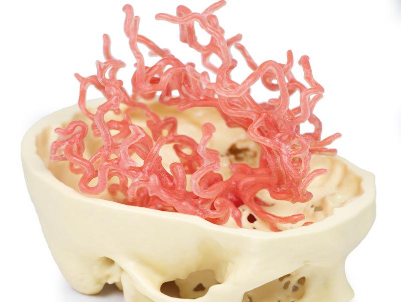
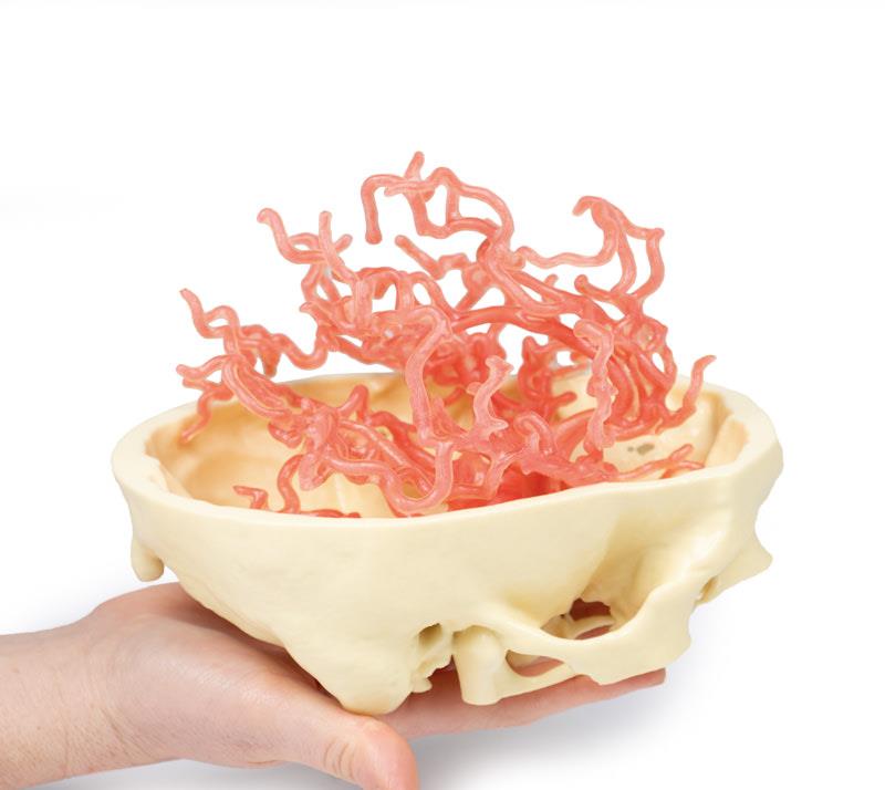
The 3D Printed Anatomy – Arterial Circulation is a detailed anatomical model made for medical educators, students, and healthcare professionals. It provides a comprehensive view of cerebral and cerebellar arterial structures for precise teaching and advanced study. Choose this product to elevate anatomical education and enhance vascular understanding with unmatched clarity.
Unlock Deep Vascular Insight with the 3D Printed Anatomy – Arterial Circulation
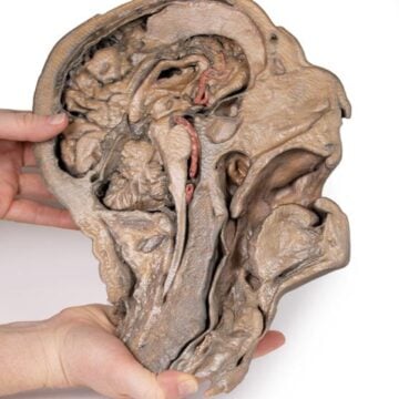
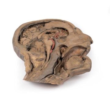
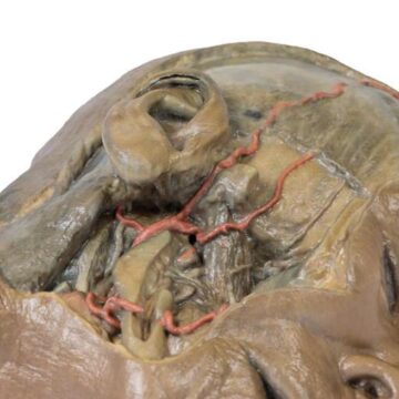
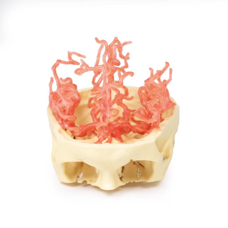
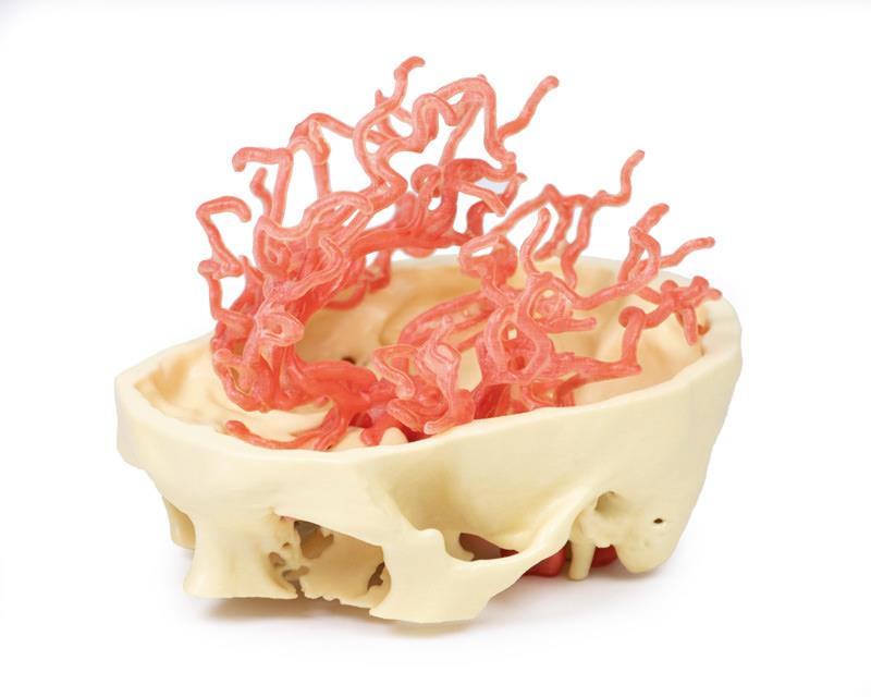
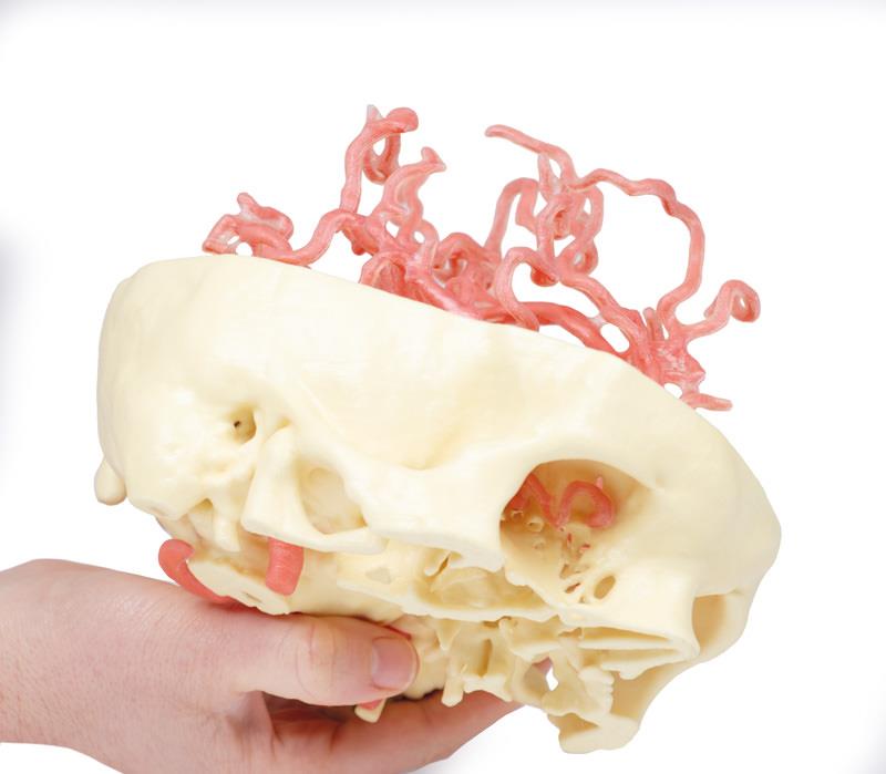
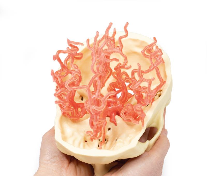
This anatomical model delivers an expanded, clinical-grade visualization of the arterial network that supplies the brain. Using advanced angiographic data and precise segmentation, it demonstrates the internal carotid and vertebral arteries as they enter the skull and branch into complex intracranial arteries. The model includes the complete Circle of Willis, branching patterns of cerebral and cerebellar arteries, and detailed features such as the pericallosal arteries, superior and inferior divisions of the middle cerebral arteries, and the posterior cerebral branches. You can easily view the ophthalmic artery as the first intracranial branch of the internal carotid entering the optic canal. This model meets education standards for clarity and accuracy, empowering both teaching and in-depth anatomical study.
Features and Benefits
- Expanded 3D-printed model based on clinical angiographic data segmentation
- Accurately displays internal carotid and vertebral arteries entering the skull
- Shows full branching of cerebral and cerebellar arteries including the Circle of Willis
- Highlights pericallosal arteries with all named branches
- Details both superior and inferior divisions of the middle cerebral artery
- Includes sulcal, temporal, and parietal arteries
- Illustrates posterior cerebral artery branches clearly
- Features the ophthalmic artery as it enters the optic canal
- Meets clinical and educational standards for anatomical accuracy
- Supports advanced teaching, demonstration, and student learning
Indications for Use
- Medical and allied health education settings
- Student study and advanced anatomy courses
- Clinical demonstration and patient education
- Reference for neurological and vascular anatomy
Size Guide
- This model is provided in a single, enlarged clinical-education scale based on actual patient angiographic data
- Dimensions closely match real human arterial structures for teaching accuracy