Features
The Pituitary Adenoma is a detailed 3D printed anatomical model designed for medical educators, students, and healthcare professionals. It provides a tactile and visual reference for understanding the pathology, diagnostic imaging, and surgical anatomy of pituitary tumors. Choose this model to enhance your clinical education, achieve precision teaching, and support informed medical decision-making.
Discover Exceptional Anatomical Clarity with the Pituitary Adenoma
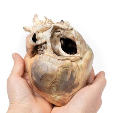
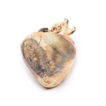
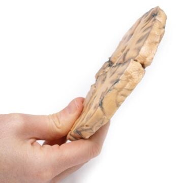
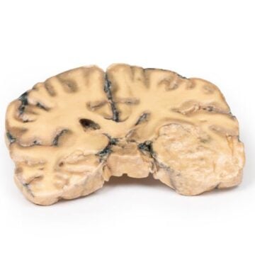
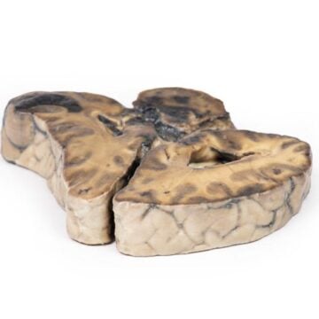
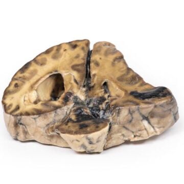
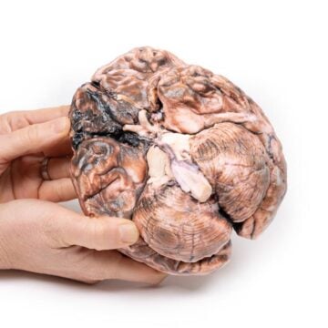
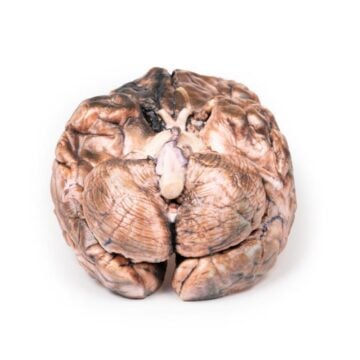
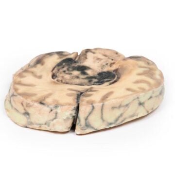
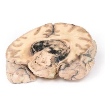
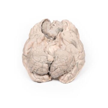
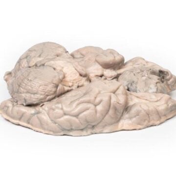
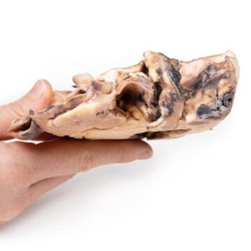
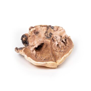
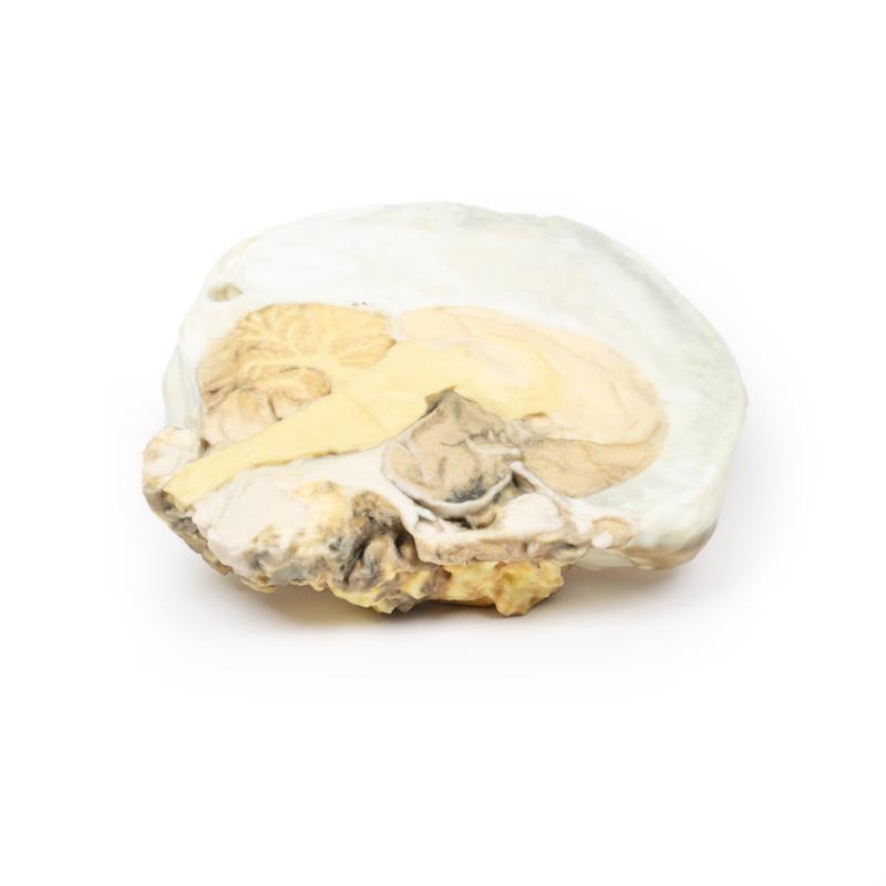
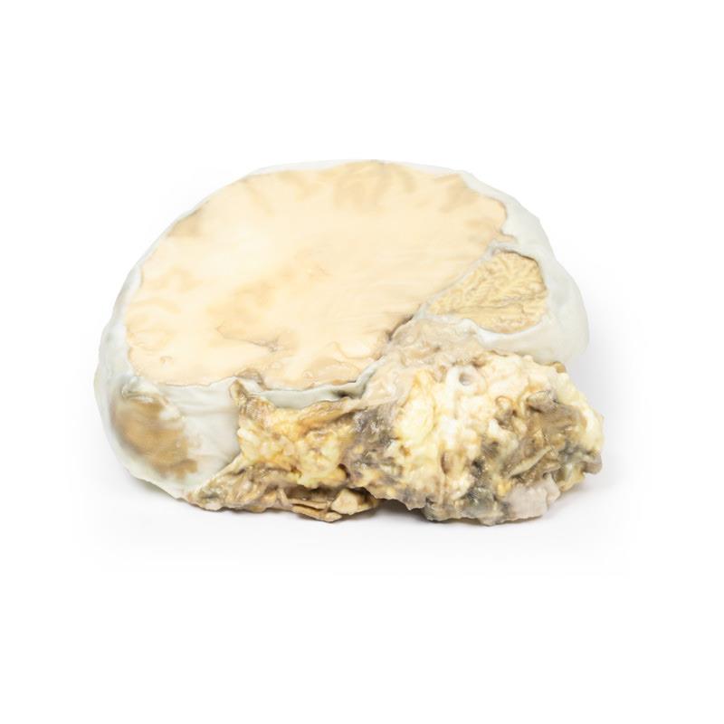
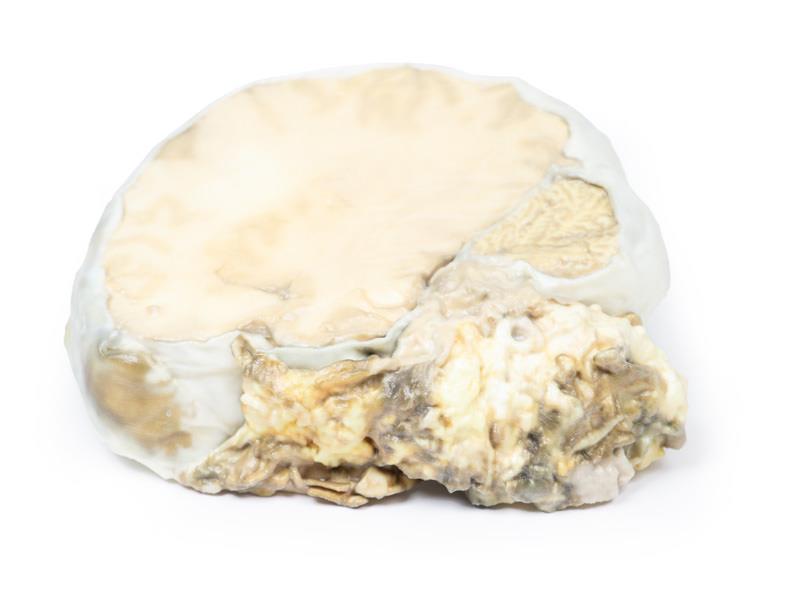
This clinical-grade 3D printed model showcases a complete replacement of the pituitary gland by a 4cm round tumor, capturing bony erosion, sphenoid bone destruction, and optic chiasma compression. The Pituitary Adenoma model visually and physically demonstrates the effects of tumor expansion, including upward midbrain displacement and sellar enlargement. Use it to teach and learn about clinical presentation, imaging findings, and surgical challenges. Its ultra-realistic pale brown homogeneous surface, precise sagittal brain slice, and inclusion of related anatomical structures offer unmatched educational value. Rely on this premium model for accurate, hands-on understanding of the most common pituitary tumor in adults.
Features and Benefits
- 3D printed clinical-grade anatomical model of a chromophobe adenoma
- Presents pituitary gland completely replaced by a 4cm tumor
- Shows sphenoid bone erosion and enlarged sella turcica
- Includes compressed optic chiasma and displaced midbrain
- Displays pale brown homogeneous tumor surface with area of hemorrhage
- Replicates sagittal brain slice to right of falx cerebri
- Ideal for education, research, and surgical planning
- Durable, precision manufacturing for repeated handling
Indications for Use
- Medical and anatomy education for students and professionals
- Patient consultation and explanation of pituitary tumors
- Demonstrating mass effect and clinical symptoms (headache, blurred vision, hemianopia)
- Teaching diagnostic imaging and surgical approach to pituitary lesions
Size Guide
- Actual anatomical dimensions with tumor – maximum diameter 4cm
- Sagittal plane specimen, includes midbrain, falx cerebri, and optic chiasma
- Model precisely represents the real-life case—refer to image for proportions