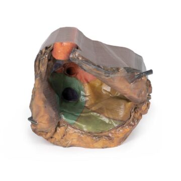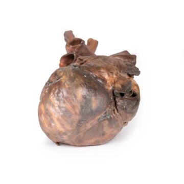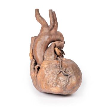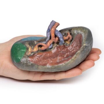

Features
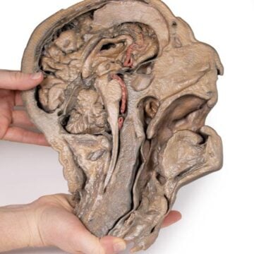
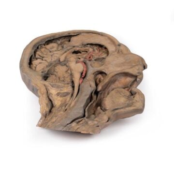
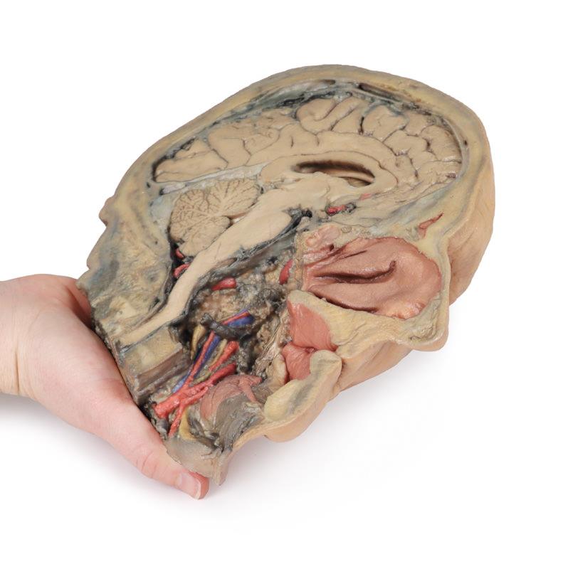
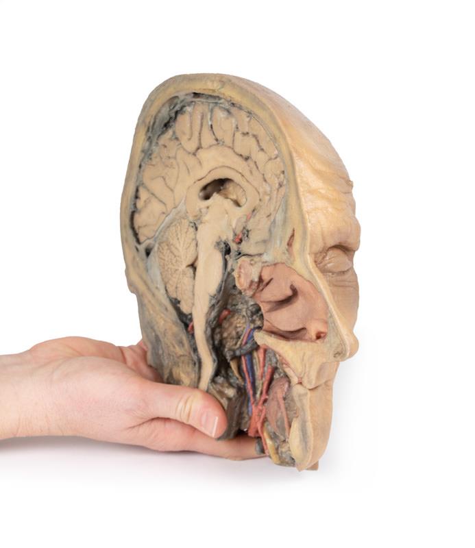
The Median Section through head sagittal section of head with deep dissection is a specialized 3D printed anatomical model designed for educators, students, and clinicians. It provides detailed visibility into midline brain and head structures along with a unique deep dissection of the pharyngeal region. This model solves the challenge of visualizing both superficial and intricate neurovascular anatomy, making it a vital teaching resource for complex head and neck anatomy.
Unlock Deep Insights into Head Anatomy with This Precision 3D Model
Enhance your anatomical teaching and study with the Median Section through head sagittal section of head with deep dissection. This clinical-grade model combines an accurate midsagittal cut of the head, preserving essential brain features and cranial cavity structures, with advanced dissection of the pharyngeal region. The printed model exposes key neurovascular and glandular structures through strategic removal of bone, constrictors, and oral mucosa, giving users unparalleled access to both common and deep anatomical landmarks. With everything from the cingulate gyrus to the ascending pharyngeal artery clearly displayed, you gain an optimal blend of clear educational value and detailed realism.
Features and Benefits
- True midsagittal section with preserved brain and cranial cavity structures
- Unique deep dissection of the pharynx via removal of basicranial bone, anterior atlas, and axis
- Preserved dura mater, tentorium cerebelli, and falx cerebelli
- View superior sagittal sinus and confluence of sinuses in cross-section
- Retains cingulate gyrus, sulcus, and lateral ventricle visibility
- Highlights diencephalon, midbrain, cerebellar hemisphere, and fourth ventricle
- Exposes both anterior cerebral artery and posterior inferior cerebellar artery (false coloured)
- Displays dissected neurovascular bundles including internal/external carotid arteries, jugular vein, vagus nerve, and sympathetic trunk
- Exposes submandibular gland, lingual artery, genioglossus, and hypoglossal nerve
- Digitally eliminates undissected side for unobstructed view
- Clinical-grade, 3D printed educational quality
Indications for Use
- Anatomical education and teaching in universities, medical schools, and allied health courses
- Patient demonstration and consultation for head, brain, or neck procedures
- Surgical planning or review for ENT, neurology, maxillofacial, and dental professionals
- Detailed study of neurovascular and glandular head structures
Size Guide
- Single 1 -1 scale model representing midsagittal and deep-dissected head anatomy
- SKU ERLMP1105
- Accurate life-size dimensions as per preserved human head anatomy