Features
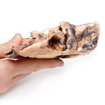
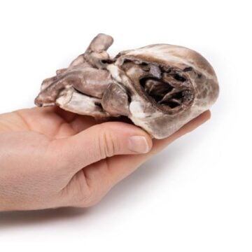
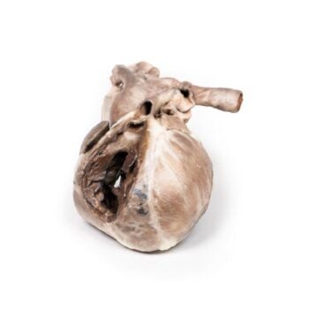
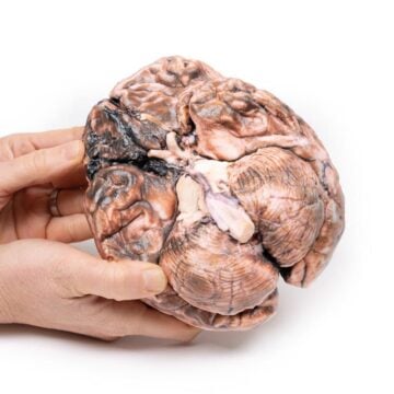
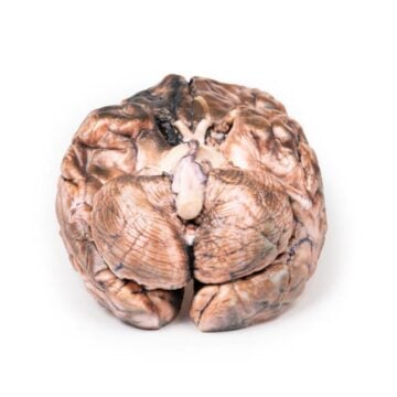
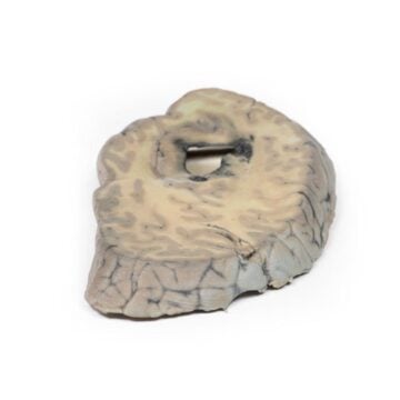
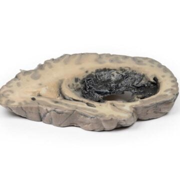
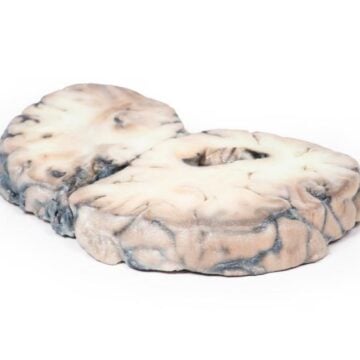
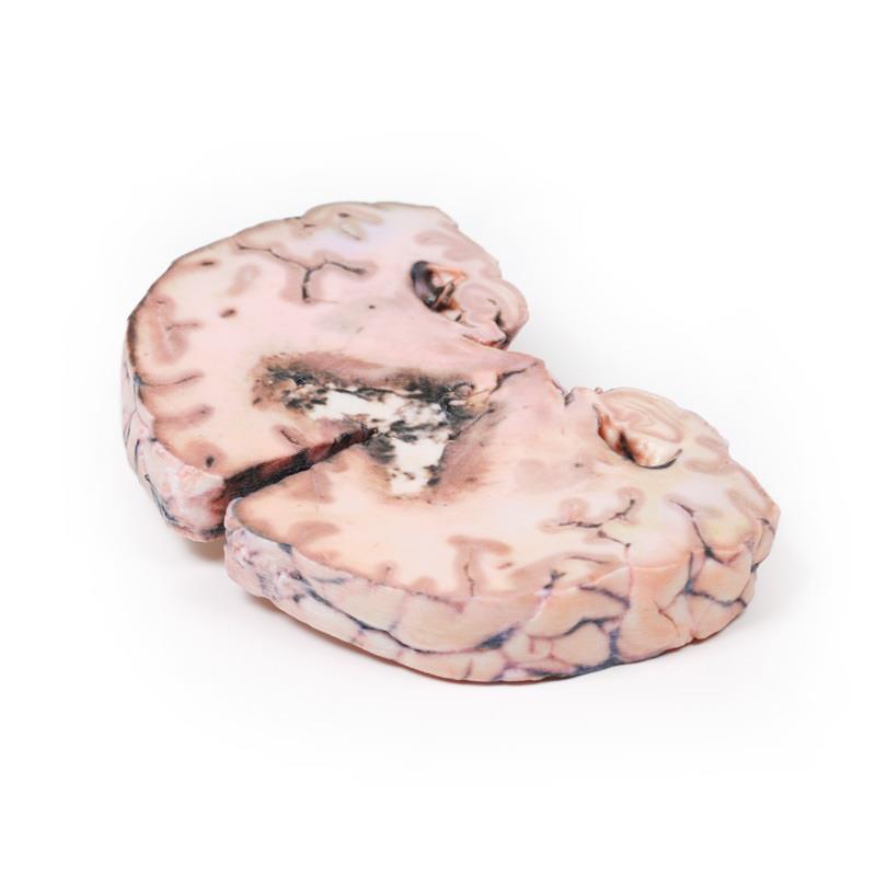
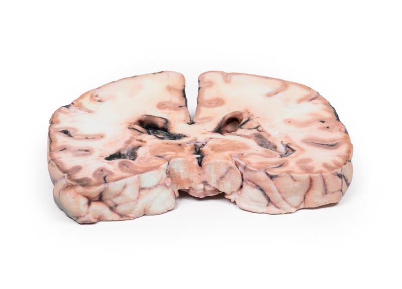
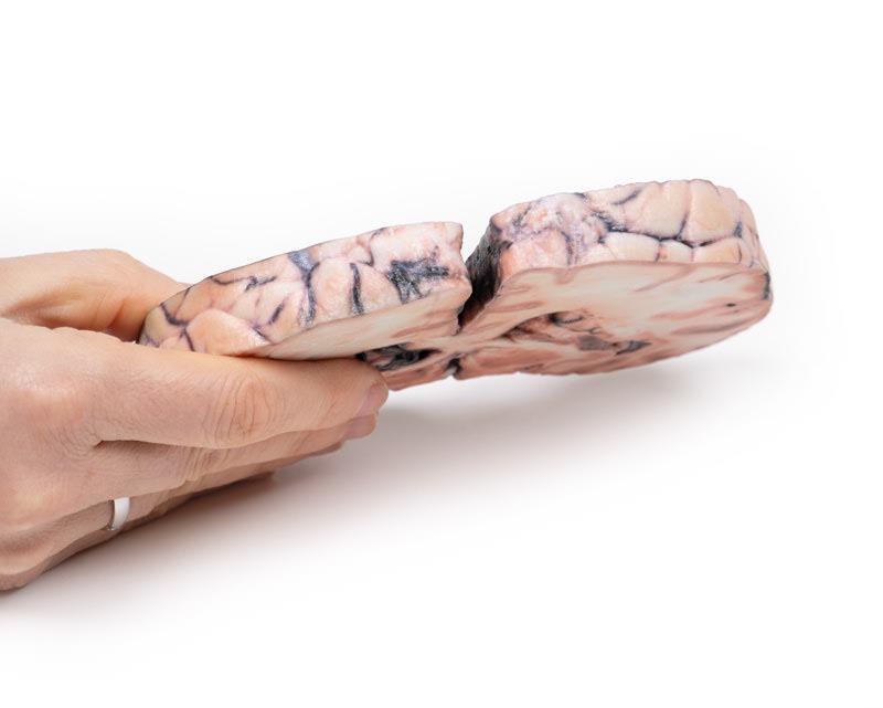
The Glioma grade 3-4, causing papilloedema is a detailed 3D printed anatomical model crafted for medical educators, students, and clinical professionals. It provides an accurate physical representation of an advanced brain tumor with associated papilloedema, giving a valuable visual and tactile resource for teaching, training, and case study discussions. Choose this model to enhance anatomical understanding and demonstrate complex pathology that textbooks alone cannot convey.
Examine Advanced Brain Pathology with the Glioma grade 3-4, causing papilloedema Model
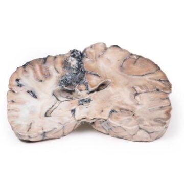
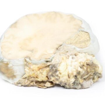
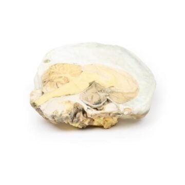
This clinical-grade model recreates a real patient’s experience with high-grade glioma and secondary papilloedema. A 24-year-old female developed seizures, blurred vision, and headaches before diagnosis. The model highlights the obliterated lateral ventricles, distortion of the aqueduct, and tumor infiltration through key brain structures. Vascular regions, haemorrhages, and necrosis produce a mottled, variegated appearance, immersing users in actual pathological anatomy. Expert medical model makers have ensured high educational value and realistic detail, making this an essential teaching asset for neurology and pathology programs.
Features and Benefits
- High-fidelity 3D printed model from real clinical case
- Displays a large, well-demarcated intracerebral lesion
- Shows obliteration of lateral ventricles and internal capsule
- Highlights infiltration across corpus callosum
- Exhibits vascularity, haemorrhage, and necrosis detail
- Educational quality suitable for teaching and demonstration
- Durable design allows repeated handling and study
- Supplied by a trusted Australian medical store
Indications for Use
- Medical and anatomy education programs
- Clinical staff training and patient education
- Student examination and practical demonstration
- Tumor pathology and neurology teaching
- Case-based learning in academic settings
Size Guide
- Full-size life-like brain section displaying advanced glioma—actual anatomical proportions
- Model represents true-to-life dimensions of a 24-year-old adult brain