Features
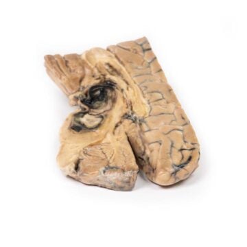
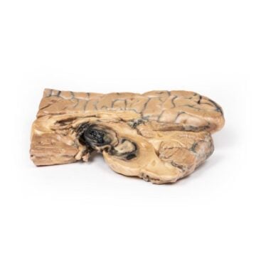
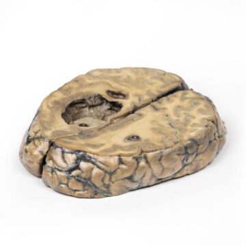
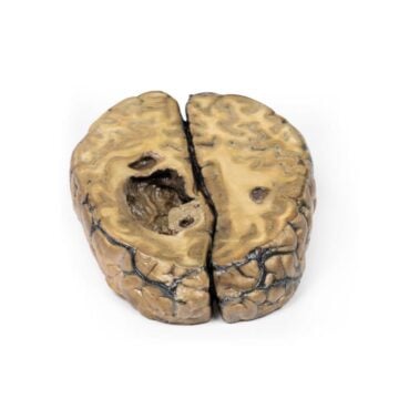
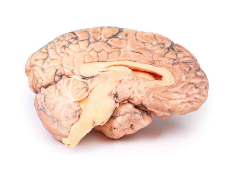
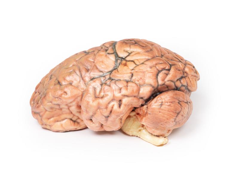
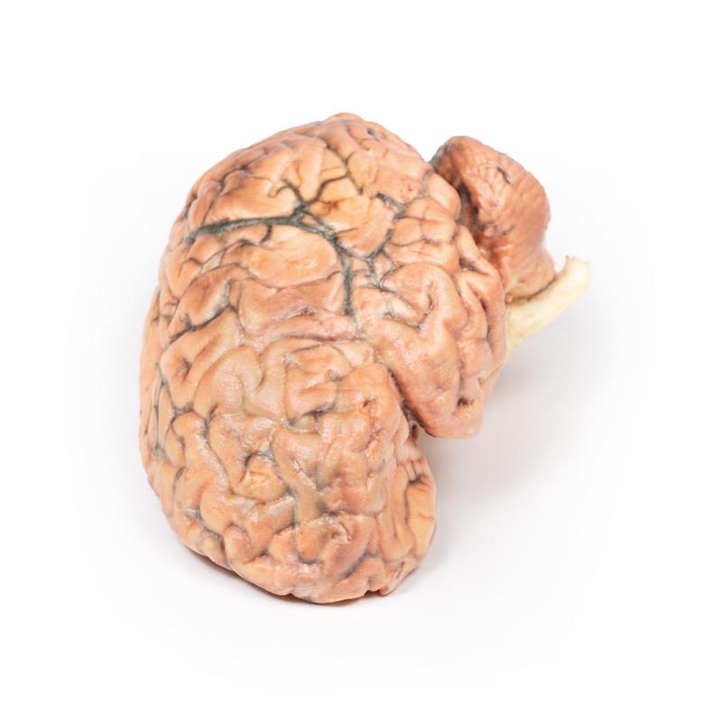
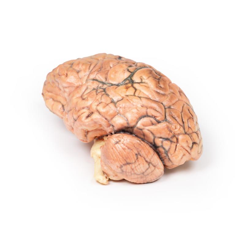
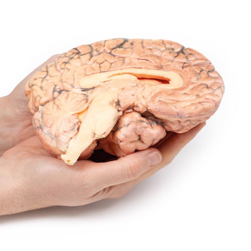
The Craniopharyngioma is a clinical-grade 3D printed brain model crafted for medical educators, students and neurosurgical teams. It delivers a detailed, realistic visualisation of this rare brain tumour, aiding in clear understanding of complex neuroanatomy and tumour pathology. This model helps facilitate deeper insight into tumour location, surgical challenges, and neuroanatomical relationships that textbooks cannot provide.
Enhance Neuroanatomy Education with a Realistic 3D Model
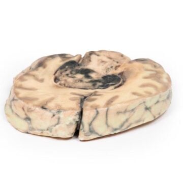
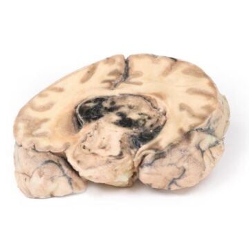
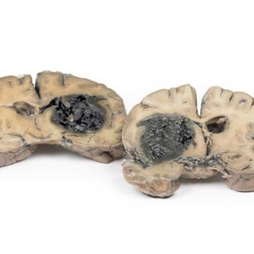
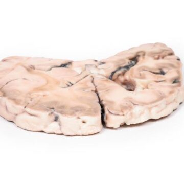
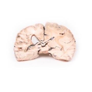
This 3D printed Craniopharyngioma model showcases a meticulously replicated sagittal section of the brain, centred on the hypothalamic region. Designed using actual patient pathology, the model displays an ovoid tumour measuring 2.5 x 1.5 cm which distorts the third ventricle and impacts surrounding critical structures. Medical professionals benefit from examining solid and microcystic tumour components, encapsulation details and the effects on the optic chiasm and ventricular system. With precise anatomical landmarks and distinguishing characteristics, this model prepares learners and professionals for both diagnosis and treatment planning.
Features and Benefits
- Clinical-grade anatomical accuracy for teaching and demonstration
- High-fidelity 3D print based on real pathological data
- Displays tumour relationship to the hypothalamus, third ventricle, and optic chiasm
- Shows capsule, ventral surgical excision site, and microcystic tumour features
- Highlights foramen of Munro obliteration and displacement of neuroanatomical structures
- Useful for pre-surgical planning and pathology review
- Durable construction for repeated educational use
Indications for Use
- Medical and pathology education
- Neurosurgical training and procedure simulation
- Patient and family education
- Demonstration of tumour impact on brain anatomy
Size Guide
- Tumour size on model 2.5 x 1.5 cm
- Sagittal brain section at life-size anatomical scale
- No additional sizing variations available