Features
The Astrocytoma is a clinical-grade 3D printed anatomical model designed for medical professionals, students, and educators. It provides an accurate, hands-on visualization of a high-grade brain tumor, supporting advanced learning in neuropathology and oncology. By choosing this model, you gain access to a precise replica of a pathology specimen that enhances both teaching and diagnostic insight.
Enhance Your Medical Training with a True-to-Life Astrocytoma Model
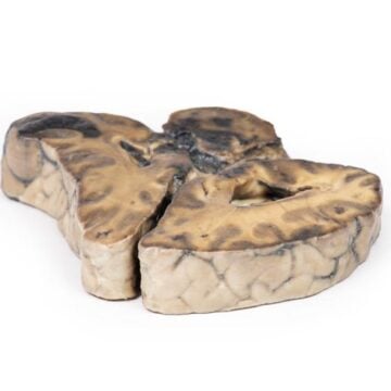
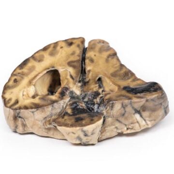
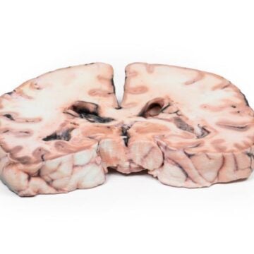
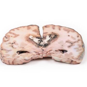
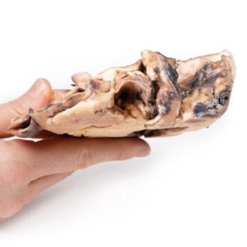
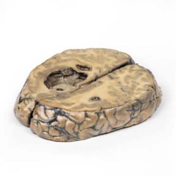
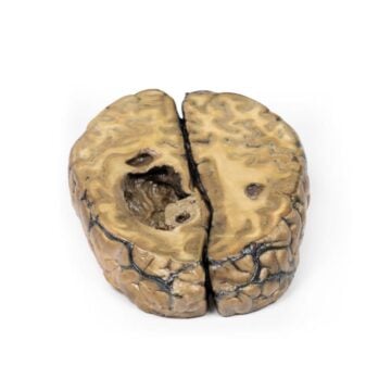
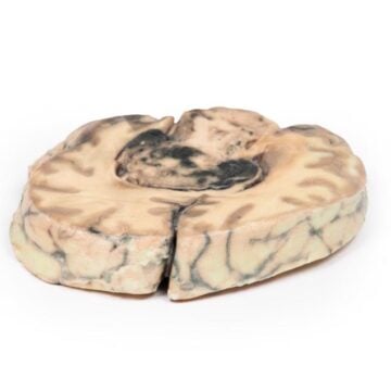
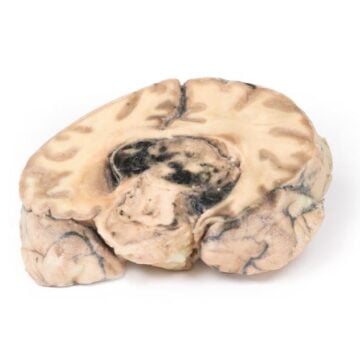
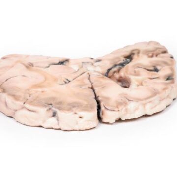
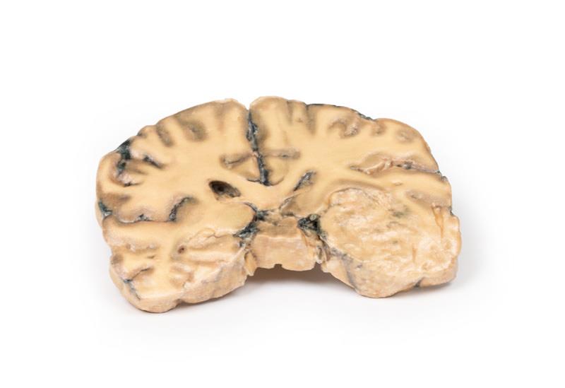
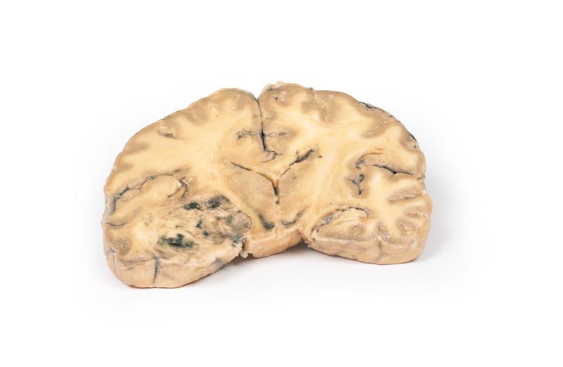
This Astrocytoma model authentically replicates a coronal section of the human brain, revealing a tumor localized in the right temporal lobe. The model shows realistic features including hemisphere enlargement, subtle gyral flattening, and visible subfalcine herniation. Hemorrhagic and necrotic regions illustrate disease progression, while fine anatomical details support clear understanding of Grade III/IV astrocytomas. Designed for education, the model demonstrates typical histological characteristics such as gemistocytes and a fibrillary matrix. Utilize this resource in pathology instruction, clinical review, or surgical planning, as it adds value that paper-based materials or illustrations cannot match.
Features and Benefits
- Replicates a real coronal brain section with a temporal lobe tumor
- Shows poorly demarcated tumor mass, hemisphere swelling, gyral pattern flattening
- Visualizes subfalcine brain herniation and advanced necrosis
- Exhibits hemorrhagic foci and differentiated zones
- Highlights anatomical and pathological tumor attributes
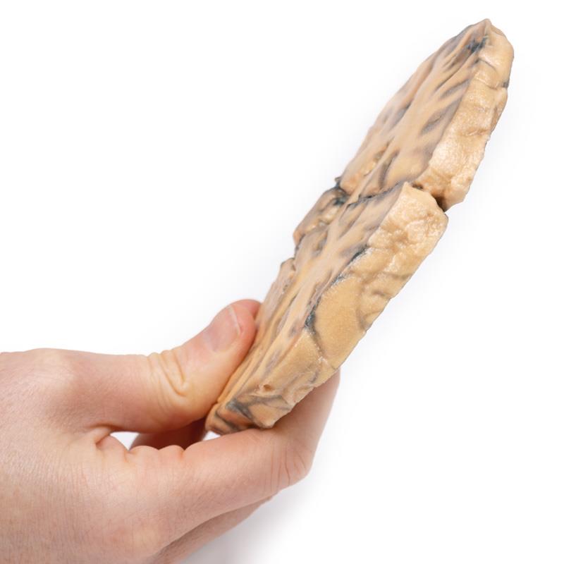
- Clinical-grade, durable 3D printed construction
- Ideal for medical education, training, and reference
Indications for Use
- Medical and pathological education on brain tumors
- Demonstrating high-grade glioma pathology
- Illustrating histological tumor features to students or trainees
- Supporting interdisciplinary tumor board review
- Patient and family education on diagnosis or treatment
Size Guide
- Life-sized model based on an actual 73-year-old female brain specimen
- Coronal brain section with right temporal lobe tumor anatomy