Features
Bring Clarity to Complex Lessons with a Realistic Fertilization Model
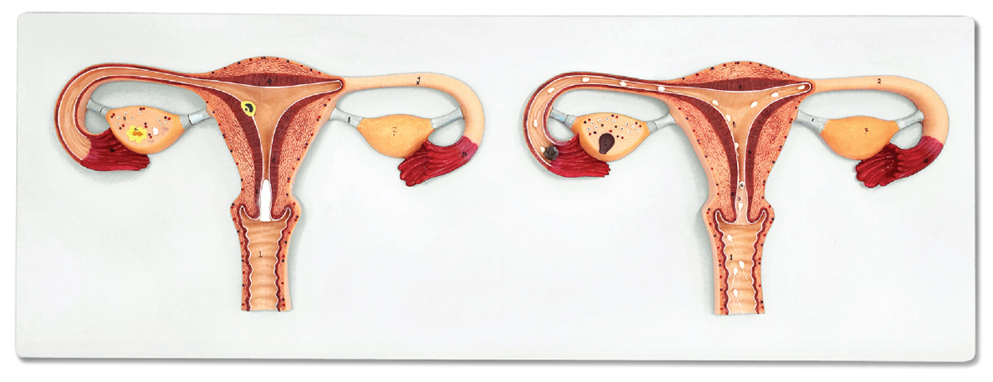
Illuminate the marvel of conception for students and trainees with the Fertilization Process Model, 2 Parts. This highly detailed anatomical teaching tool helps educators present every step of human fertilization effectively. The Fertilization Process Model, 2 Parts displays the delicate process from ovum penetration to the union of genetic material in vibrant, color-coded lifelike detail. Each durable part was molded for repeated use in classrooms, labs, and clinical training. You can separate the model to reveal clear internal structures, allowing for interactive discussion and hands-on exploration. Students benefit from close-up visuals that simplify cell functions and interactions. Teachers can rely on this professional model to inspire, engage, and provide crucial anatomical knowledge, making abstract concepts concrete and memorable.
Features and Benefits
- Shows detailed ovum and sperm fusion in two distinct parts
- Accurate anatomical structures for effective teaching
- Color-coded elements for quick learning and comprehension
- Durable material withstands frequent classroom use
- Model separates to demonstrate internal cells and developmental stages
- Suited for group instruction, exams, and interactive coursework
- Ideal for biology, medicine, and health education
Indications for Use
- Human fertilization education
- Classroom and laboratory demonstrations
- Medical and nursing school instruction
- Patient and family education
Size Guide
- Full model height: approximately 20 cm
- Each section width: approximately 10 cm
- Two detachable parts for step-by-step demonstration