
Features
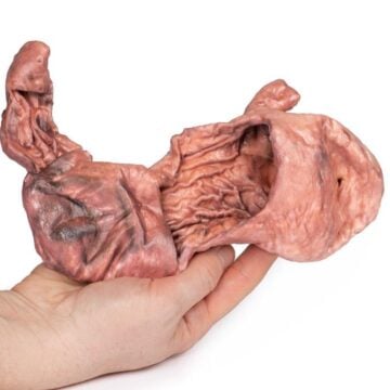
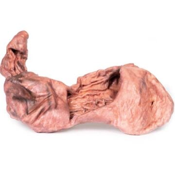
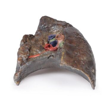
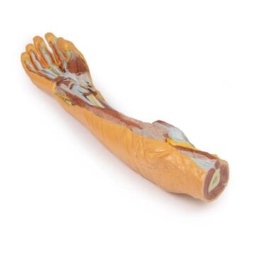
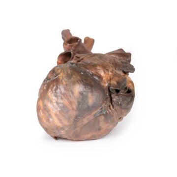
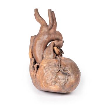
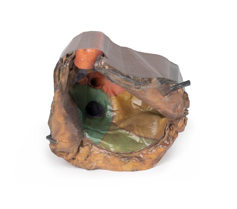
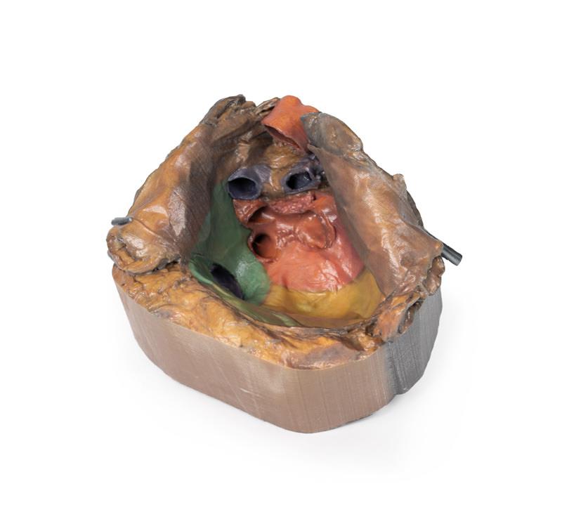
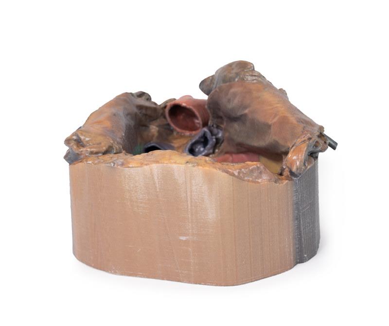
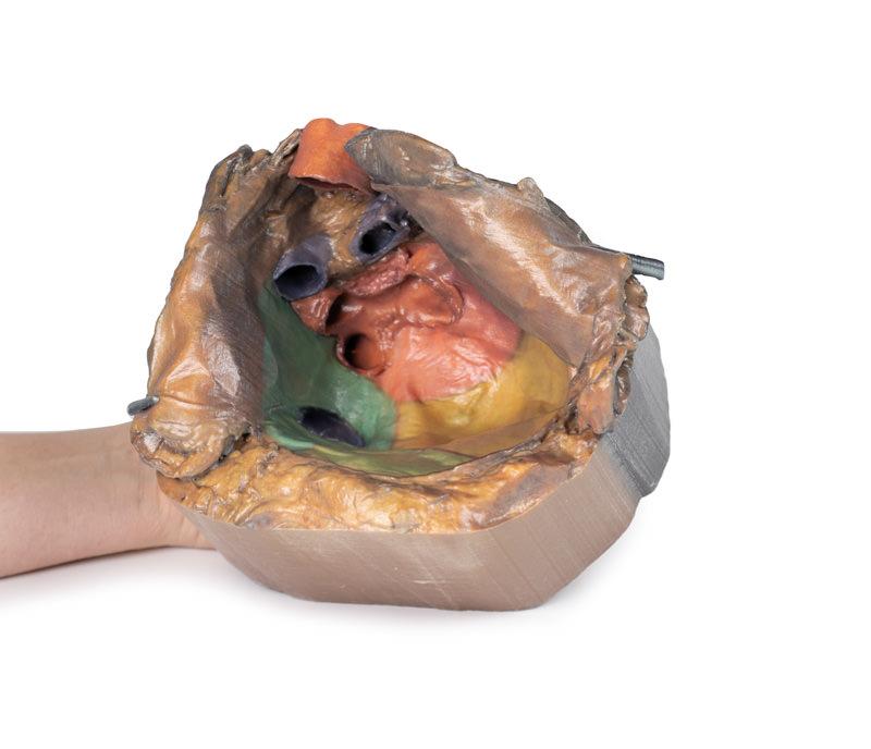
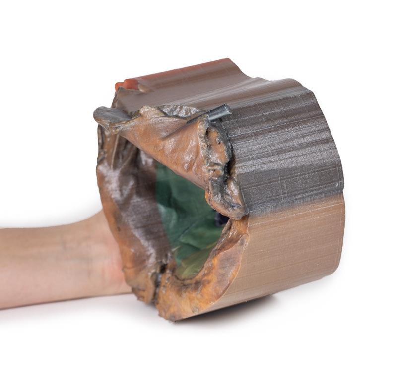
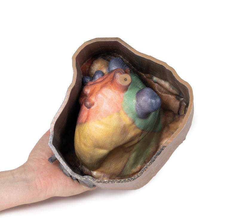
The Pericardial space is a highly detailed, clinical-grade 3D printed anatomical model designed for medical educators, students, and healthcare professionals. It offers a clear visual of pericardial reflections and mediastinal relationships by presenting the heart’s spatial orientation after the removal of the organ. This model simplifies complex anatomical structures, making it an essential teaching and study tool for advanced cardiac anatomy.
Enhance Cardiac Anatomy Learning with the Pericardial space 3D Model
This model delivers unparalleled insight into the pericardial cavity and its surrounding anatomy. By removing the heart, the Pericardial space model showcases the parietal pericardium, orientation to the diaphragm and lungs, and color-coded internal surfaces for precise regional identification. Key mediastinal landmarks, including pericardial sinuses and vascular reflections, become immediately apparent for students and clinicians. Clinical-grade detail supports accurate explanations of surgical and diagnostic approaches. Crafted using durable 3D-printed materials, this specimen withstands extensive educational use in classrooms, labs, or at the point of care. It bridges textbook knowledge with real-world visualization, ensuring advanced understanding of spatial cardiac anatomy.
Features and Benefits
- Precise 3D printed model of pericardial space with heart removed
- Showcases parietal pericardium, diaphragmatic and pulmonary surfaces
- False-colored regions highlight mediastinal anatomical boundaries
- Displays base, apex, and broad lateral (pulmonary) surfaces of the heart’s impression
- Identifies fixed and reflected pericardial regions at great vessel origins
- Highlights transverse and oblique pericardial sinuses
- Superior, posterior, anterior, and lateral orientation markers included
- Demonstrates great vessel pathways including aorta, SVC, IVC, pulmonary arteries and veins
- Detailed color-coding aids learning and instruction
- Clinical-grade durability for regular classroom or professional use
Indications for Use
- Medical and anatomy education
- Surgical and diagnostic training
- Advanced cardiac anatomy demonstration
- Patient education regarding pericardial and mediastinal relationships
Size Guide
- Model SKU – ERLMP1121
- Full-scale anatomical accuracy based on adult human thorax
- Dimensions and proportions match clinical mediastinal landmarks