Features
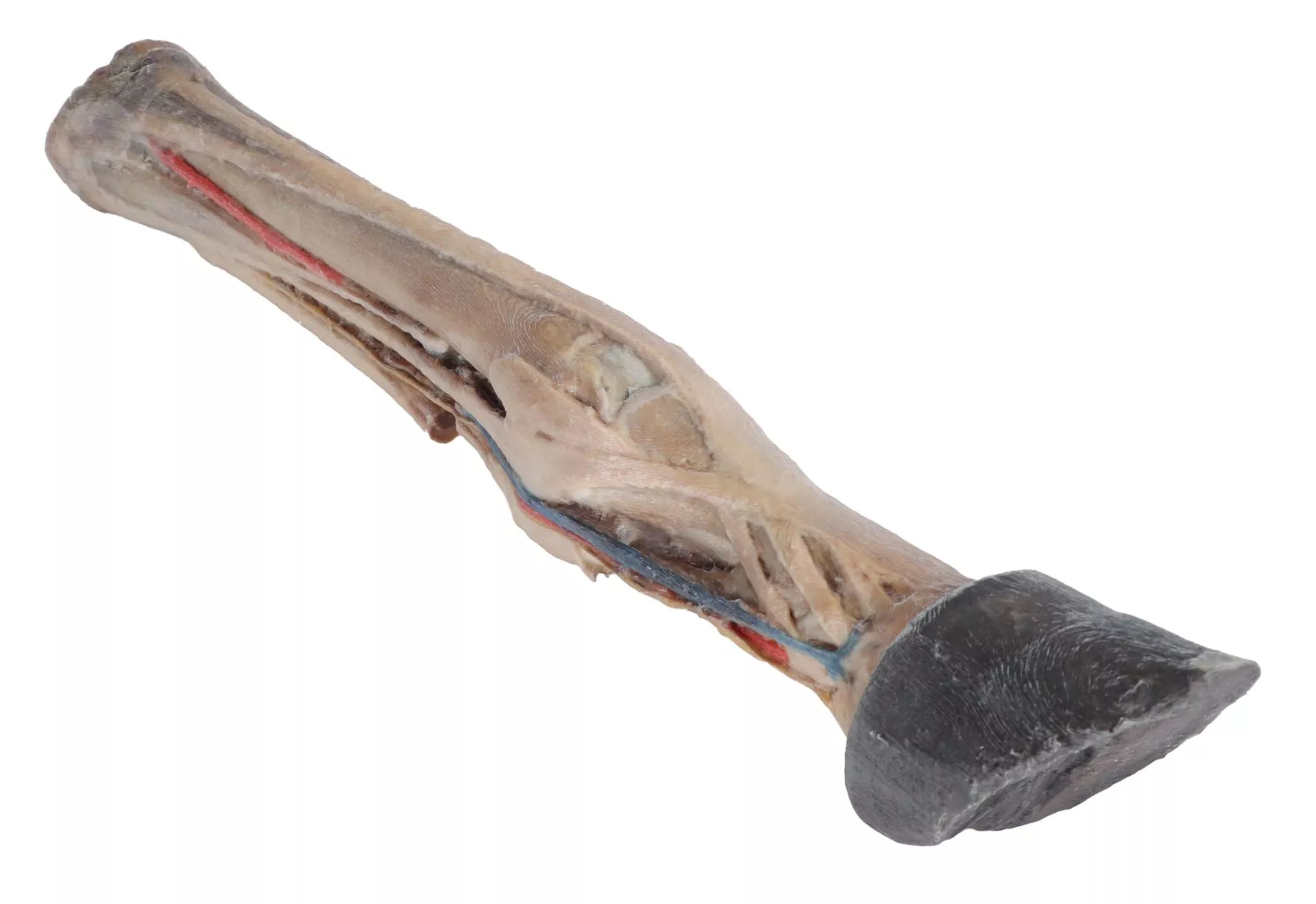
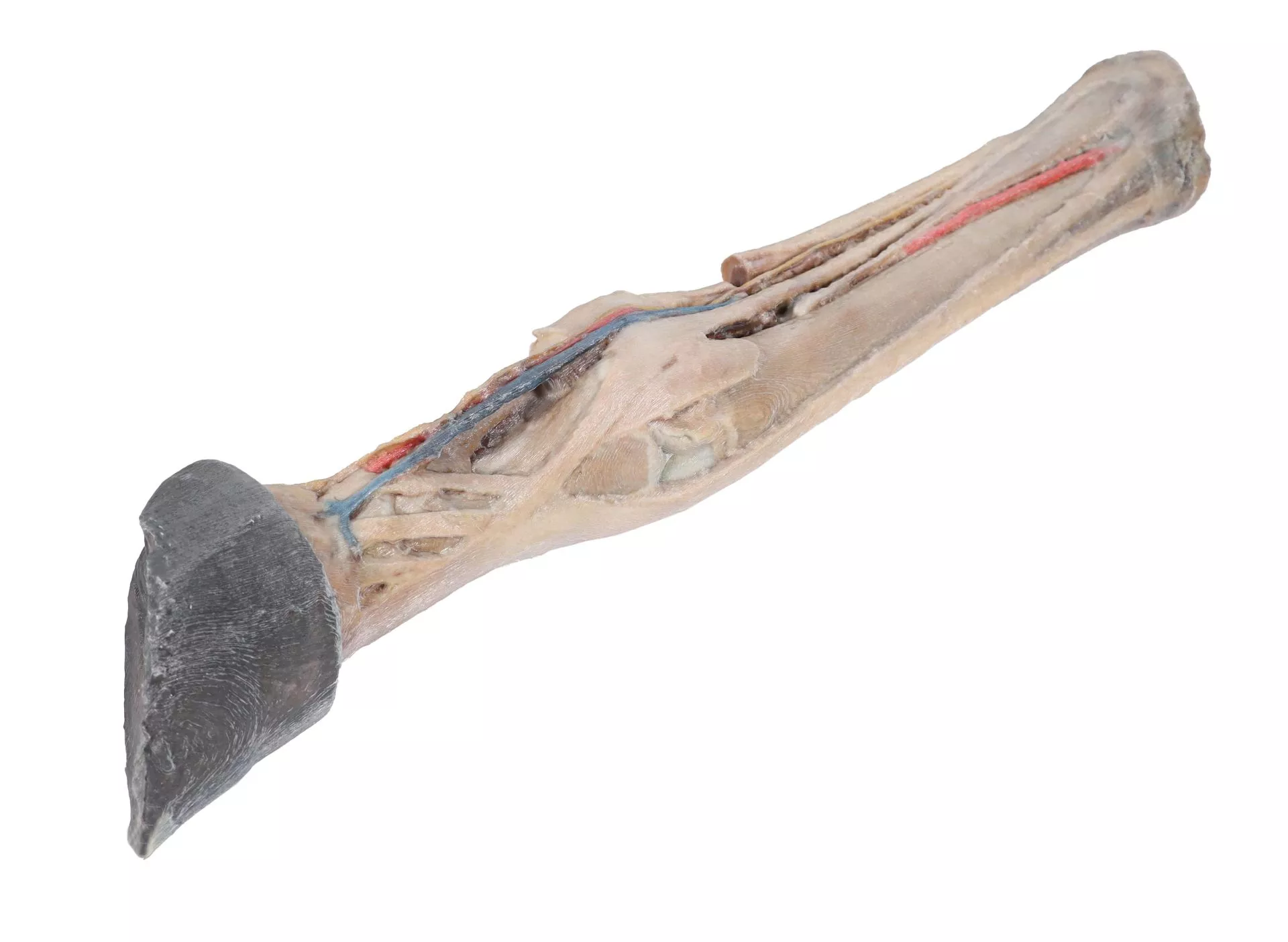
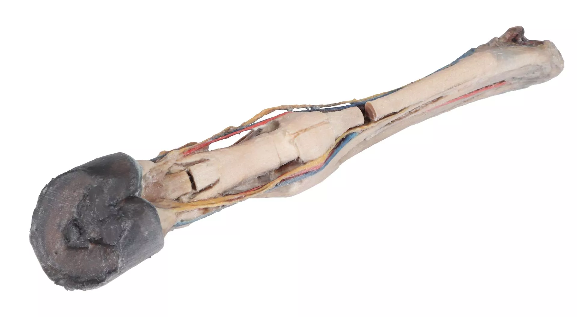
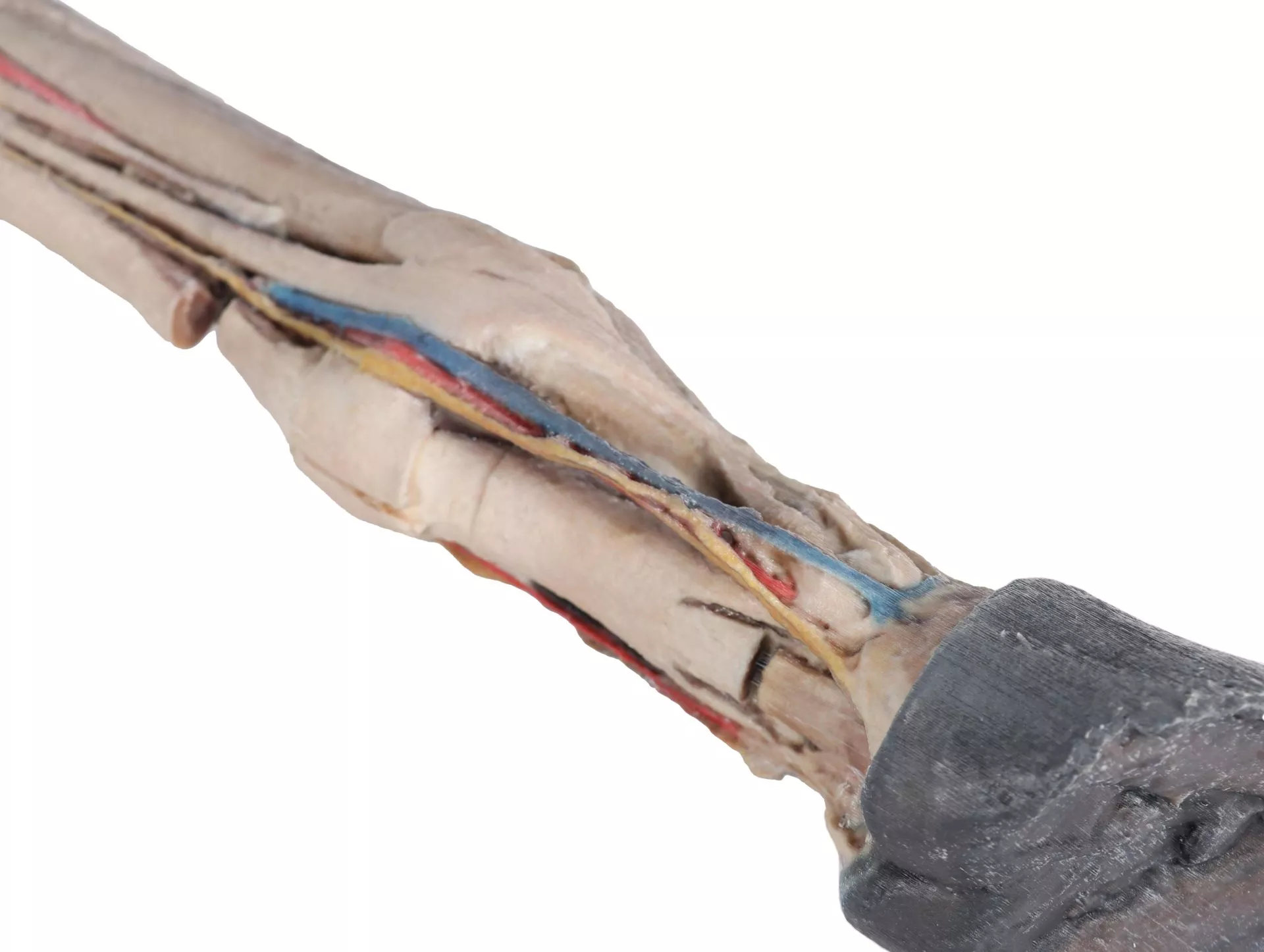
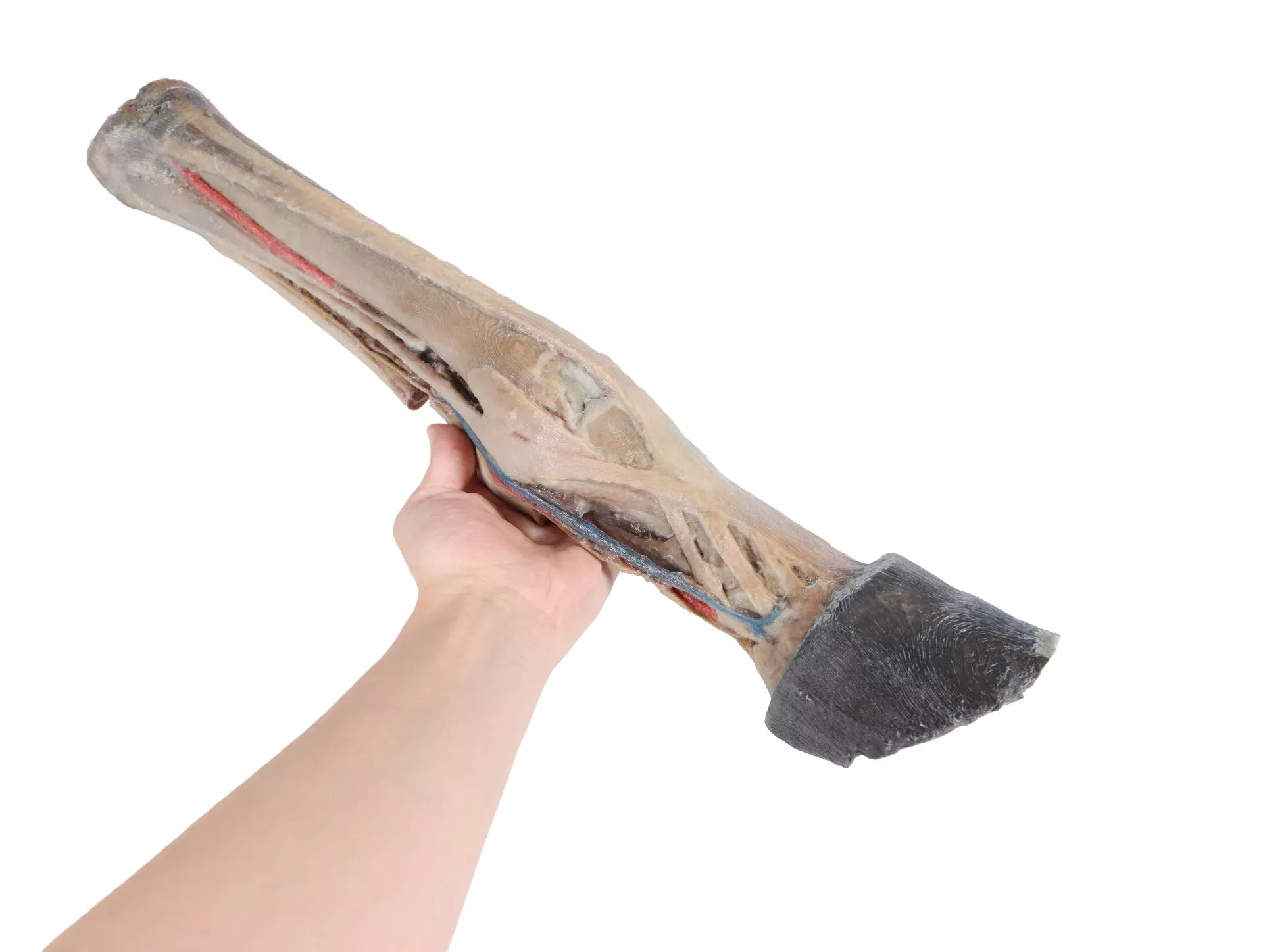
The Horse foot – tendons, ligaments, vessels and nerves – 1/3 life-size is an anatomical model created for veterinary educators, students, and professionals. It displays crucial details of equine foot anatomy in a compact, dissected specimen. Choose this model to enhance your teaching or learning with a hands-on representation that covers all vascular, nervous, and ligamentous structures.
Elevate Equine Anatomy Learning with a Detailed 1/3 Life-Size Horse Foot Model
This comprehensive specimen showcases expertly dissected equine anatomy, offering clarity on the relationship between tendons, ligaments, vessels, and nerves. The Horse foot – tendons, ligaments, vessels and nerves – 1/3 life-size enables precise study of the right foot, assisting with identification and understanding of intricate structures. Educators and students gain an exceptional tool designed for clinical-grade accuracy and durability. Unlock clear visualisation to advance study sessions and practical demonstrations.
Features and Benefits
- Displays a dissected right-foot equine specimen
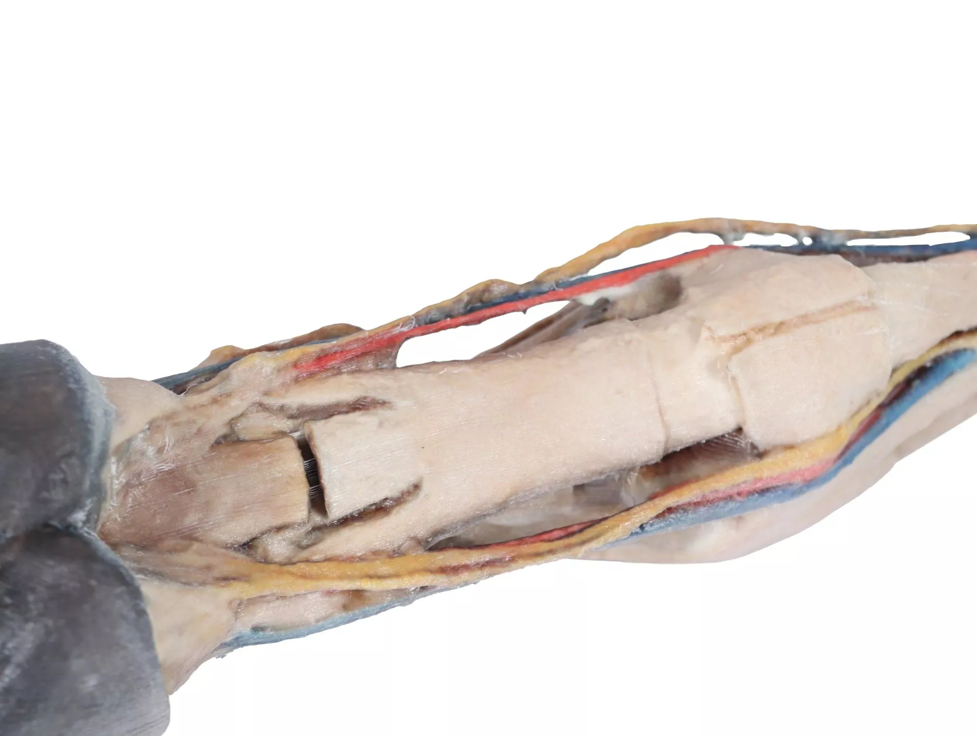
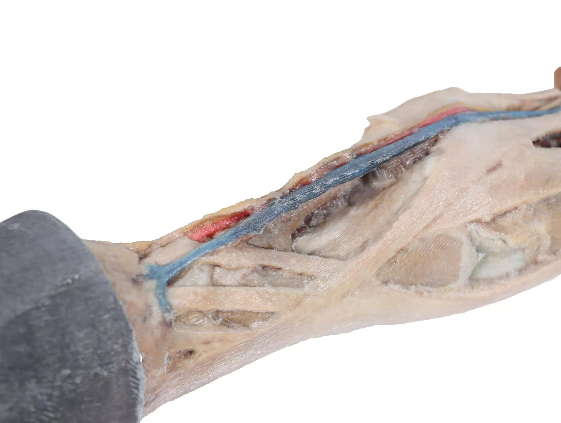
- Reveals vascular, nervous, ligament, and tendon structures
- Shows plantar digital neurovascular bundles on both sides of the flexor tendons
- Highlights the two digital extensor tendons in the proximal dorsal region
- Exposes all ligaments of the metatarsophalangeal and proximal interphalangeal joints
- Includes interosseus ligament with its sesamoid and extensor branches
- 1/3 life-size format ideal for portability and clarity
- Clinical-grade quality ensures educational and professional reliability
Indications for Use
- Veterinary medical education and training
- Professional anatomical demonstrations
- Practical study aid for students and instructors
- Reference for clinical or classroom settings
Size Guide
- 1/3 life-size anatomical specimen
- Represents a dissected right horse foot in scaled-down form