Features
Product Description
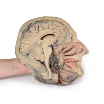
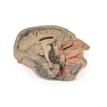
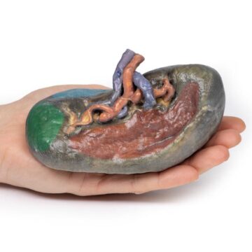
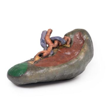
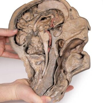
This 3D printed Superficial and deep dissection of distal leg and foot preserves a mixed superficial and deep dissection of a left distal leg and foot. Posteriorly, the compartment muscles and neurovascular structures have been removed to isolate the tendocalcaneous and expose the body of the calcaneus. Medially, the tibialis posterior and flexor digitorum longus tendons are visible deep to the crural fascia, joined by the tendon of the flexor hallucis longus as the tendons passing deep to the flexor retinaculum (opened to demonstrate the tendon passage) to the medial foot. The adductor hallucis, medial head of the flexor hallucis brevis, and flexor digitorum brevis muscles are all exposed on the medial aspect of the foot.
On the dorsum of the foot, both superior and inferior extensor retinacula are preserved, with the muscles of the anterior compartment of the leg extending to their distal attachments (including the fibularis tertius). The anterior tibial artery is exposed through to the dorsalis pedis artery. Deep to these long tendons are the extensor hallucis brevis and extensor digitorum brevis muscles, as well as the dorsal interosseous muscles. On the lateral aspect, both fibularis longus and brevis are visible deep to the crural fascia, with their tendons passing deep to both superior and inferior fibular retinacula. On the lateral margin of the foot the abductor digit minimi muscle is exposed.
Made to Order
Please Note: Thanks to the flexibility of manufacturing that 3D Printing offers, this Superficial and deep dissection of distal leg and foot model is “printed to order”, and is not typically available for immediate shipment. Most models are printed within 15 working days and arrive within 3-5 weeks of ordering, and once an order is submitted to us, it cannot be canceled or altered. Please contact us if you have specific a specific delivery date requirement, and we will do our best to deliver the model by your target date.
Advantages of 3D Printed Anatomical Models
- 3D printed anatomical models are the most anatomically accurate examples of human anatomy because they are based on real human specimens.
- Avoid the ethical complications and complex handling, storage, and documentation requirements with 3D printed models when compared to human cadaveric specimens.
- 3D printed anatomy models are far less expensive than real human cadaveric specimens.
- Reproducibility and consistency allow for standardization of education and faster availability of models when you need them.
- Customization options are available for specific applications or educational needs. Enlargement, highlighting of specific anatomical structures, cutaway views, and more are just some of the customizations available.
View the full range of 3D Printed Anatomical Models here