
Features
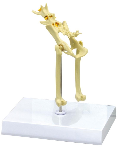
The Feline Pelvis is a detailed anatomical model designed for veterinary professionals, educators, and students. It provides a hands-on solution for studying feline pelvic anatomy and illustrates the pathology of degenerative hip osteoarthritis. Choose this model to bring real-world accuracy to teaching and demonstration, ensuring a deeper understanding of the feline musculoskeletal system.
Enhance Veterinary Learning with the Feline Pelvis Model
Deepen your anatomical knowledge with this average size Feline Pelvis model. Carefully crafted for clinical teaching and demonstration, this model features the pelvis, femur, sixth and seventh lumbar vertebrae, caudal vertebrae (tail segment), and visible spinal cord. Prominently included osteophytes represent bone spurs that result from degenerative osteoarthritis, letting you display both normal and pathological structures. Ideal for veterinary courses or clinics, it supports interactive learning and clinical education, providing realism in every detail.
Features and Benefits
- Clinical-grade quality ensures accuracy and durability
- Includes femur, L6 and L7 vertebrae, caudal (tail) vertebrae, and spinal cord
- Displays osteophytes for clear visualization of degenerative osteoarthritis in the hip joint
- Supports veterinary anatomy teaching and professional development
- Average feline size provides true-to-life scale for realistic study
Indications for Use
- Veterinary anatomy education and demonstration
- Demonstrating degenerative orthopedic conditions such as osteoarthritis
- Clinical training for students and professionals
- Educational display in teaching institutions or clinics
Size Guide
- Dimensions 18 cm length x 11 cm width x 6.5 cm height (average feline)