Features
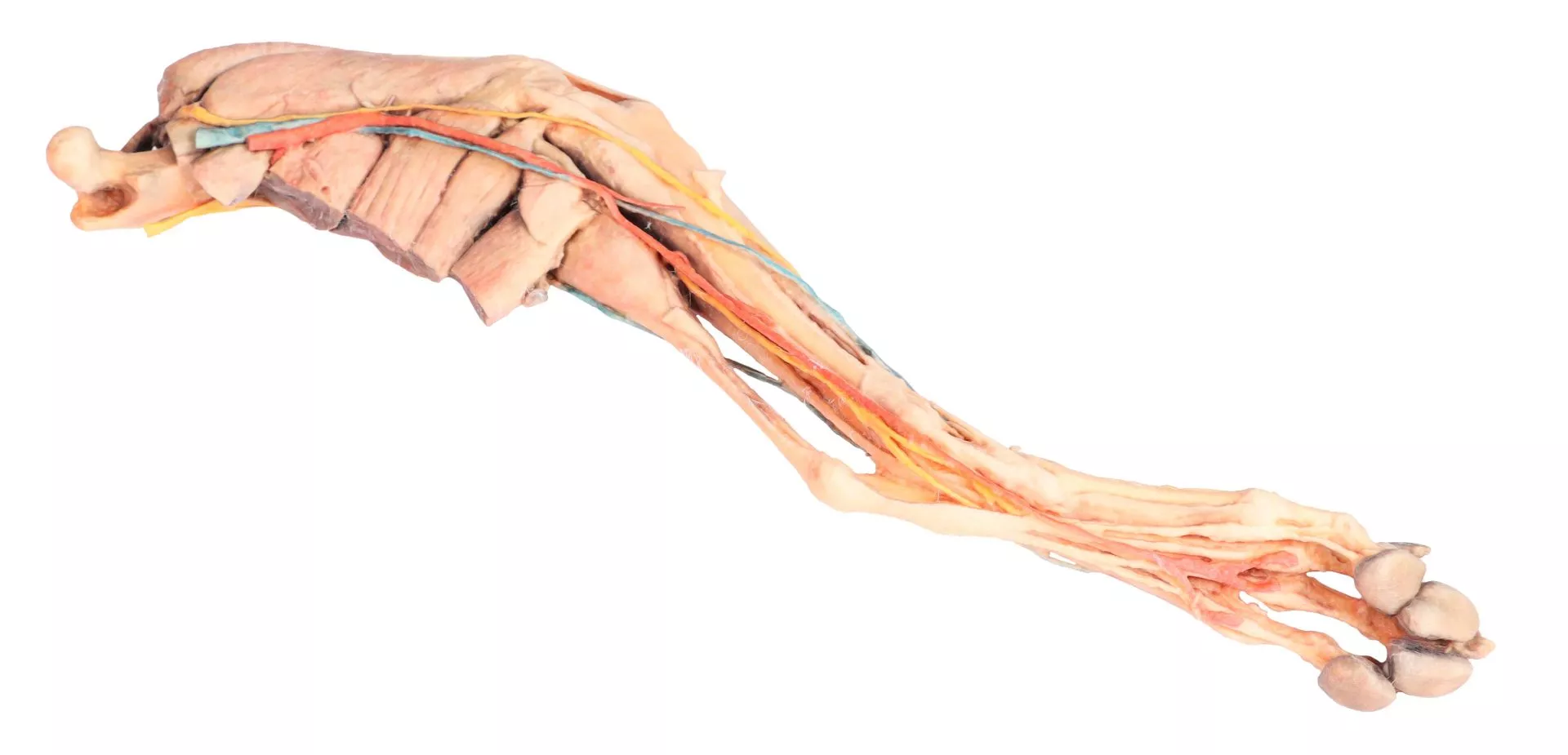
The Dog hindlimb – muscles, tendons, ligaments, vessels and nerves distal to the stifle is a clinical-grade anatomical specimen intended for veterinary educators, students, and professionals. It provides an unparalleled view of canine lower limb anatomy, helping users master essential musculoskeletal, vascular, and nervous structures for accurate instruction and confident clinical practice. This product stands out for its meticulous dissection and comprehensive anatomical exposure, making it the preferred choice for in-depth study and demonstration.
Reveal Every Critical Structure in Canine Lower Limb Dissection
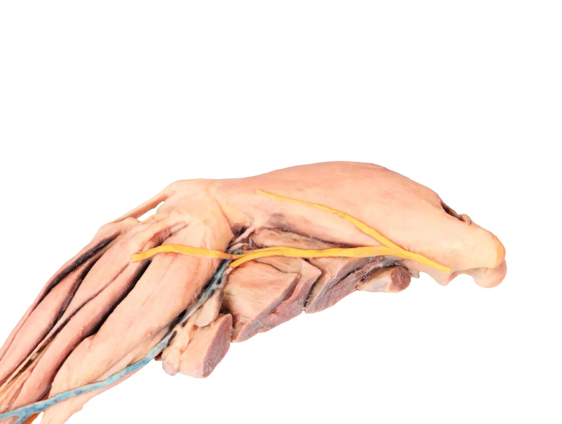
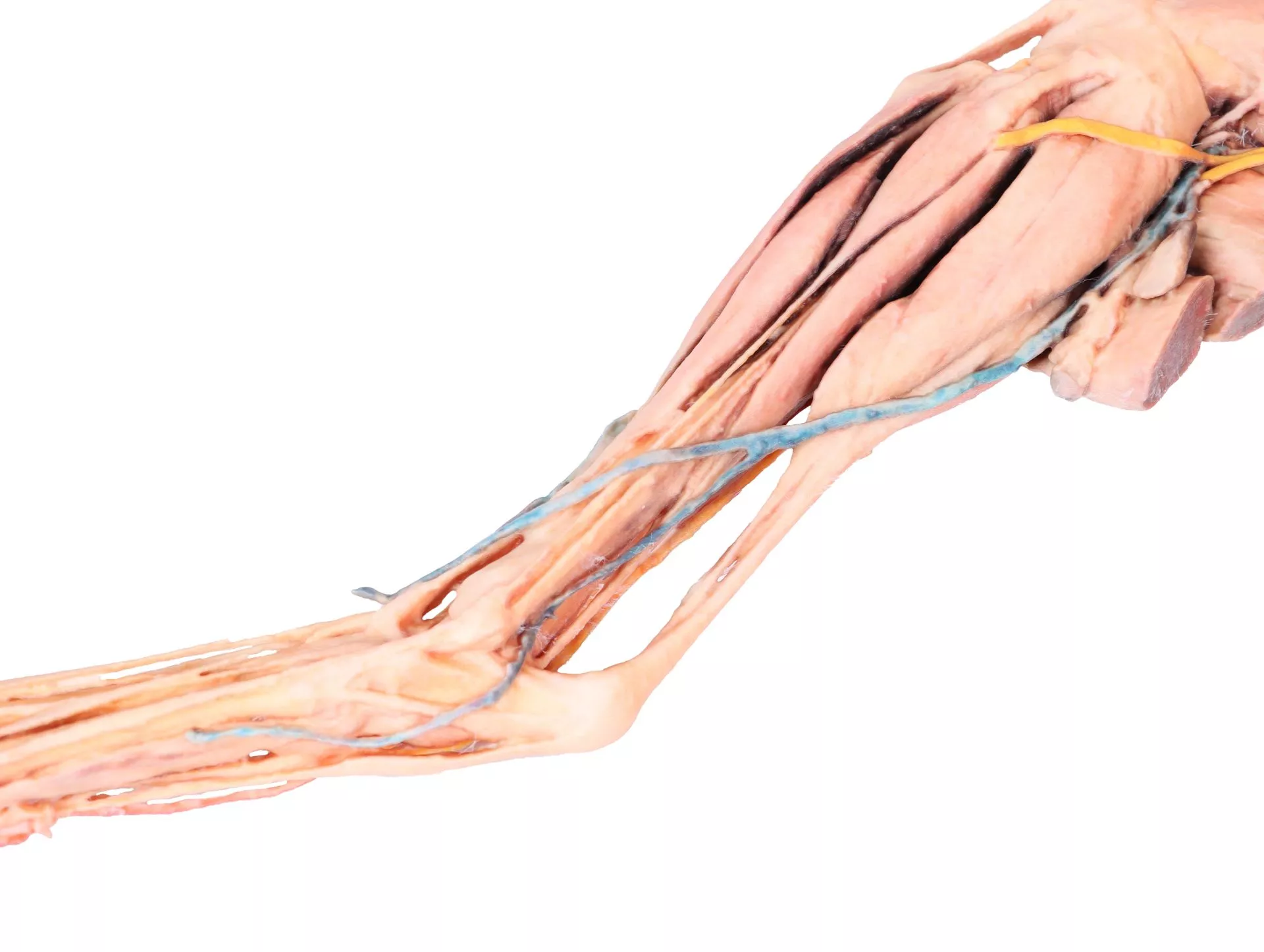
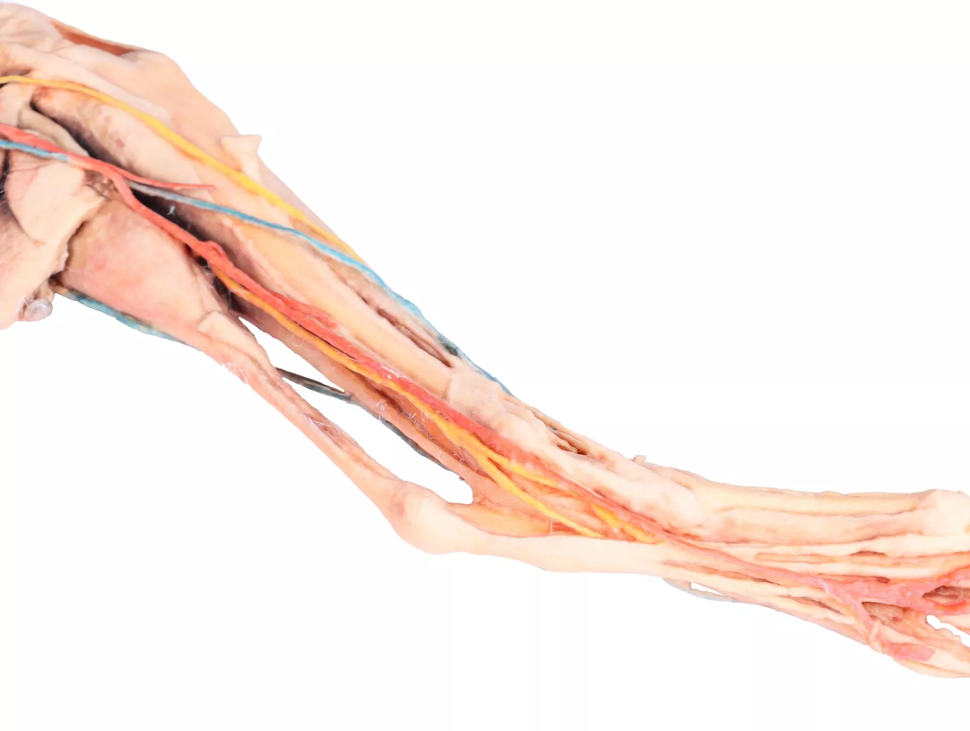
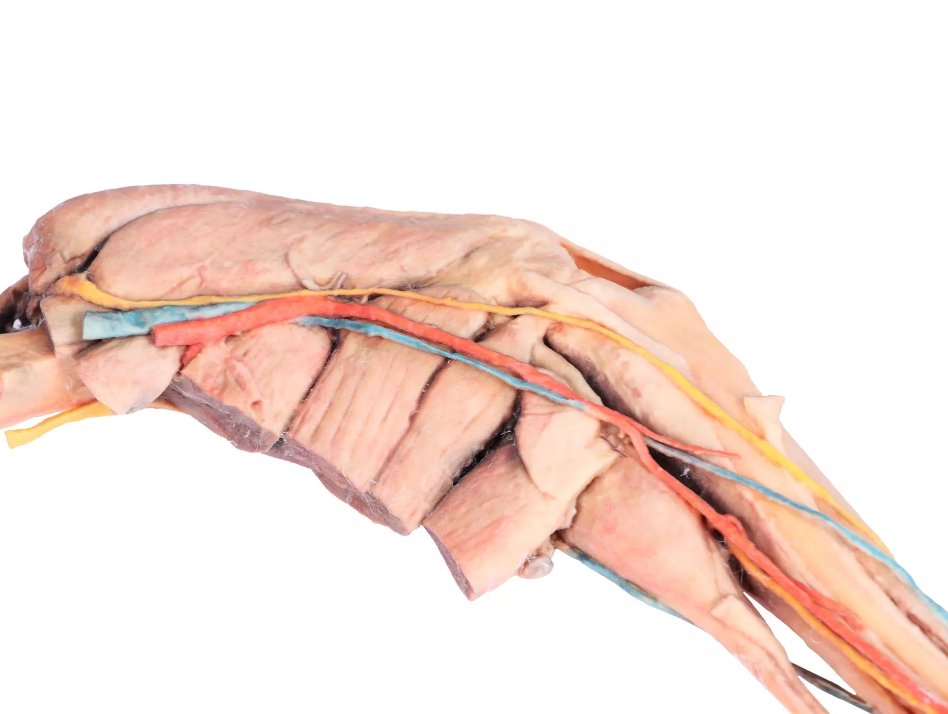
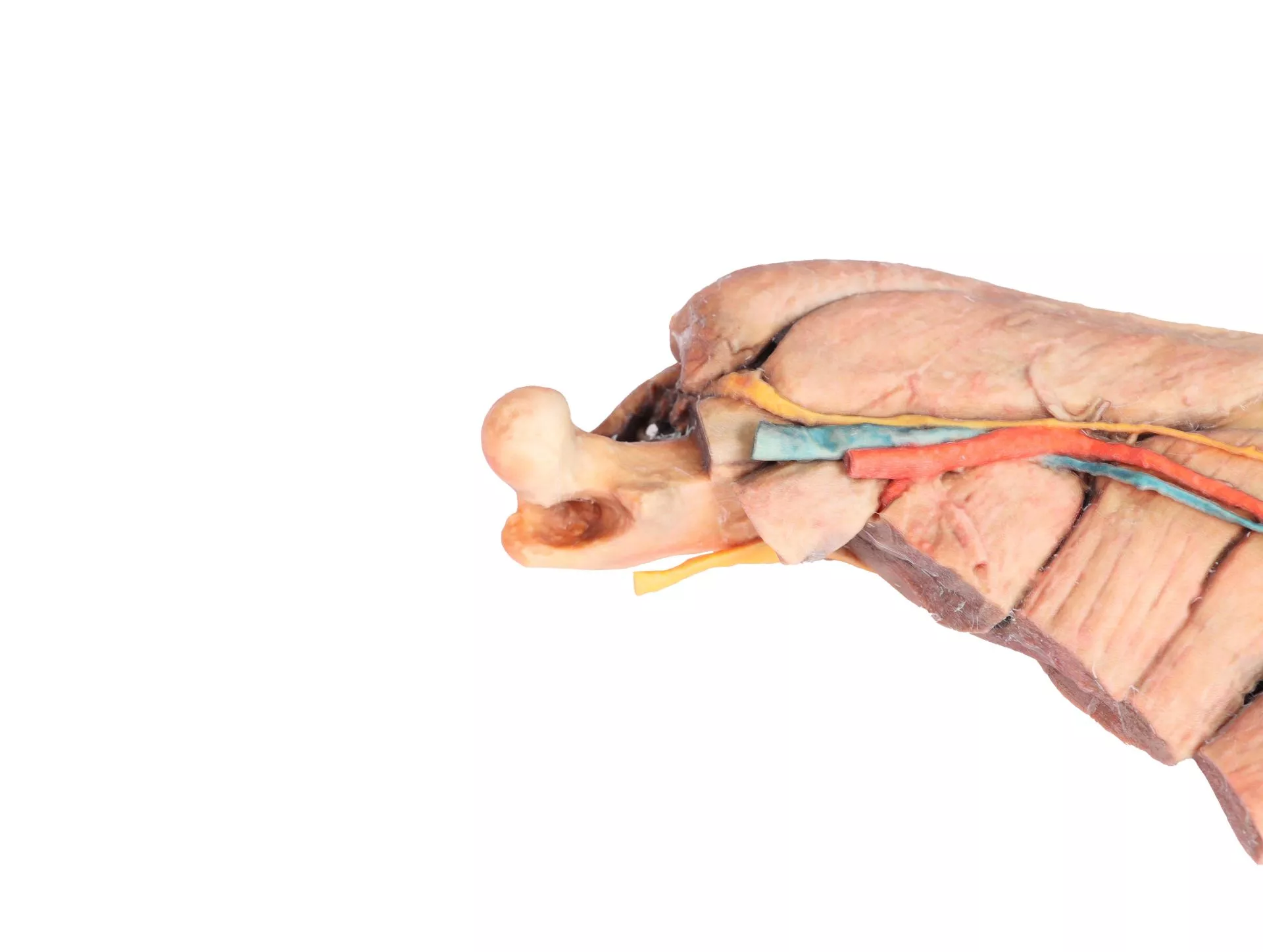
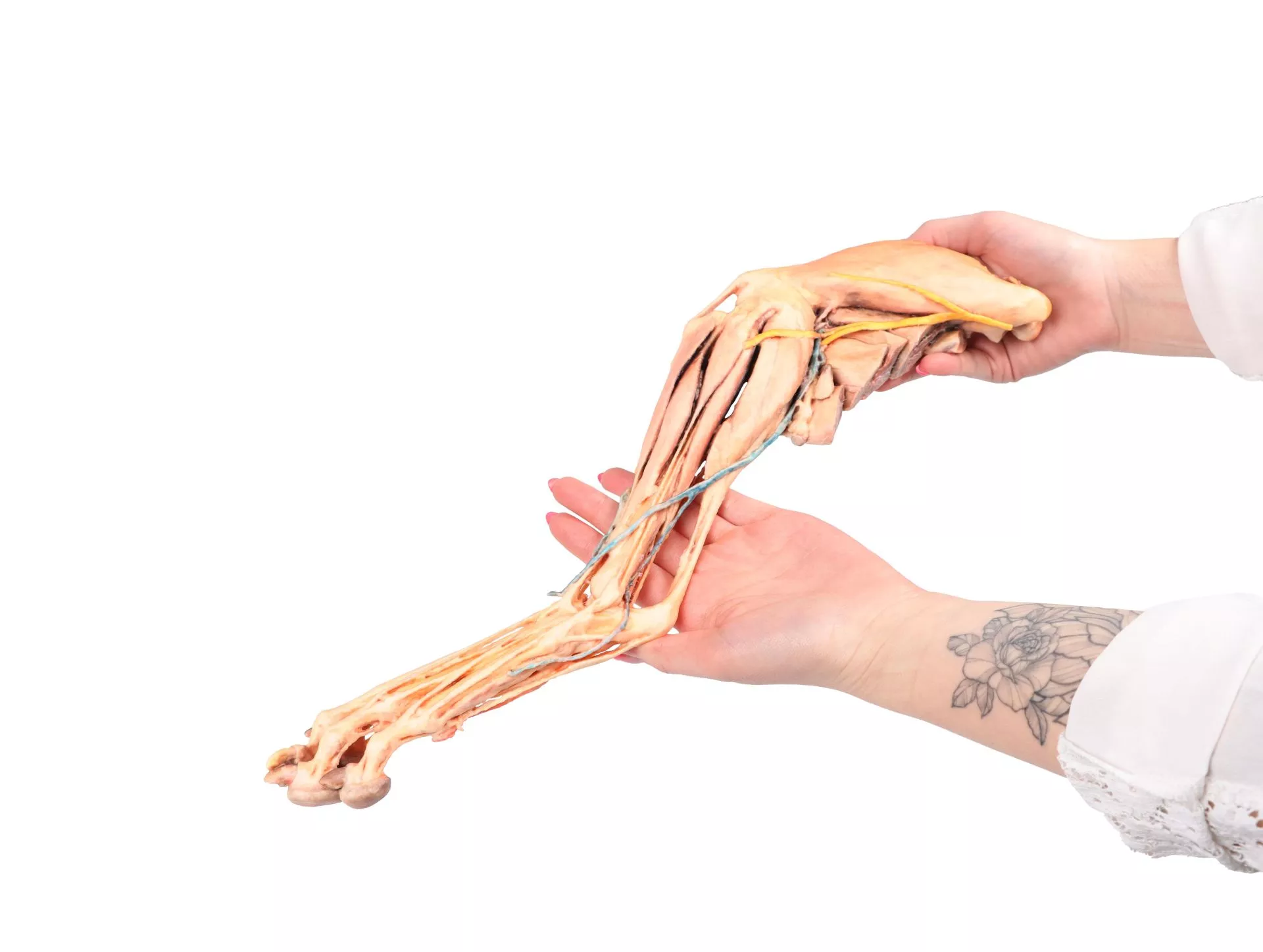
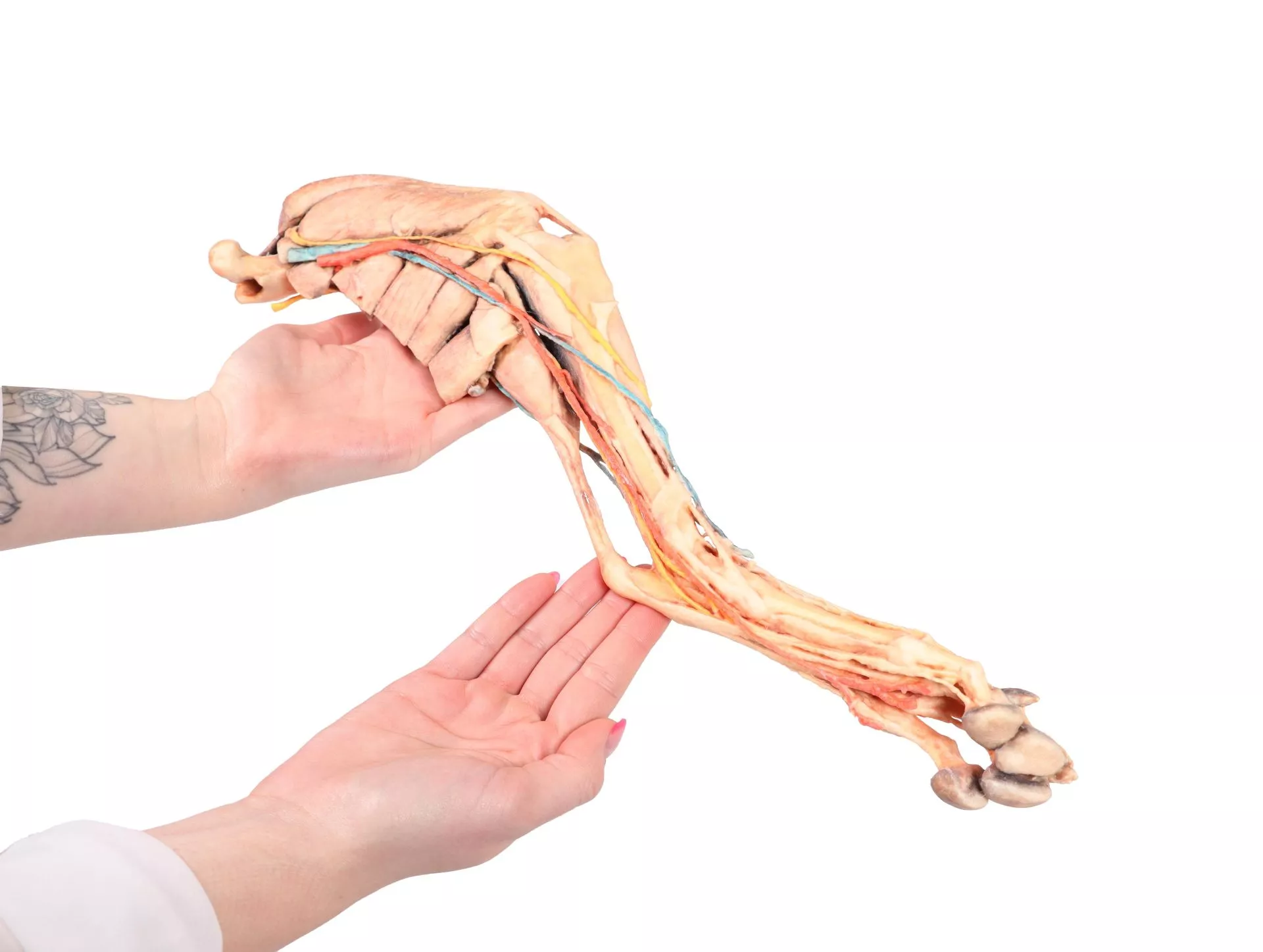
Elevate your teaching and learning experience with the Dog hindlimb – muscles, tendons, ligaments, vessels and nerves distal to the stifle. This expertly prepared specimen showcases the musculoskeletal system, tendons, ligaments, nerves, and vessels from mid-thigh to the foot. You can observe the relationships between thigh muscle insertions, a fully opened stifle joint with exposed menisci and collateral ligaments, and the detailed paths of major nerves and blood vessels. The dissection presents a clear view of extensor and flexor groupings, as well as the dorsal and palmar foot structures. Each specimen meets rigorous clinical and education standards, ensuring accuracy for practical demonstration, reference, or examination preparation.
Features and Benefits
- Superficially dissected from mid-thigh to foot
- Clearly shows thigh muscle insertions at the stifle and tibial bone
- Open stifle joint with exposed menisci and collateral ligaments
- Preserves topography of lateral saphenous vein and caudal surface
- Displays extensor and flexor muscles of tarsus and toes
- Dorsal and palmar foot structures are visible
- Includes major nerves and vessels of the femoral canal and popliteal fossa
- Detailed anatomical description available upon request
- Clinical-grade quality for educational use
Indications for Use
- Veterinary anatomy education and dissection courses
- Professional veterinary training and demonstration
- Exam preparation for veterinary students and clinicians
- Reference for surgery, diagnostics, or clinical procedures
Size Guide
- Single preserved specimen displaying mid-thigh to foot
- Includes all key structures from the stifle joint distally
- Dimensions and sizing depend on natural canine limb proportions