Features
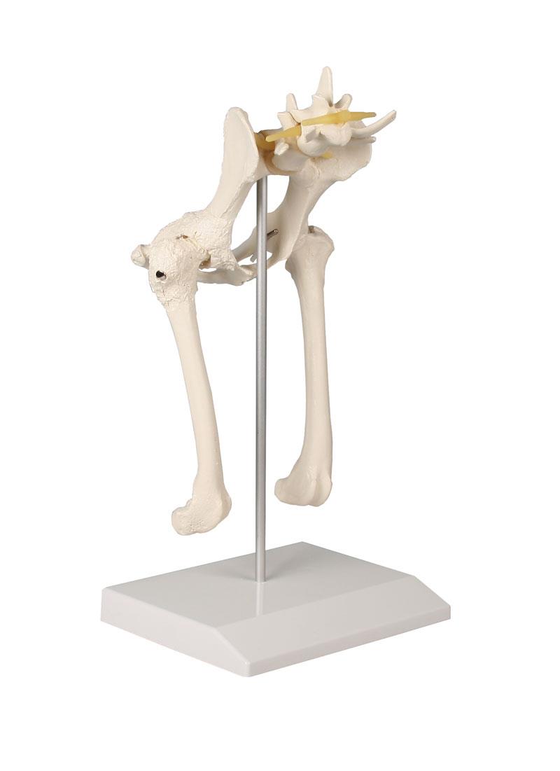
The Canine Hip is a detailed anatomical model representing an average size dog pelvis for veterinary professionals, educators, and students. It demonstrates both normal and osteoarthritic bone structures, making it an essential tool for learning about canine hip anatomy and joint disease. This model helps users visualize key structures and provides clarity when explaining complex conditions.
Discover Clinical-Grade Canine Anatomy with the Canine Hip
Enhance your understanding of veterinary orthopedic health with this precise Canine Hip model. You see the contrast between healthy and osteoarthritic bone, offering insights during lectures, demonstrations, or case reviews. The model displays the body of ilium, greater trochanter, femoral head in acetabulum, herniated disc, femoral neck, nerve pathways, the sacrum, and third trochanter. Clinical-grade craftsmanship ensures every anatomical landmark stands out, supporting detailed study and discussion in both educational and clinical environments.
Features and Benefits
- Shows normal and osteoarthritic bone structures for clear comparison
- Displays detailed anatomy including body of ilium, greater trochanter, head in acetabulum, herniated disc, and neck of femur
- Includes nerve pathways, sacrum, and third trochanter for comprehensive study
- Durable, clinical-grade construction lasting through repeated hands-on learning
- Created for veterinary education and clinical reference
- Generous size at 18 x 14 x 28 cm for effective demonstration
Indications for Use
- Teaching canine hip anatomy to veterinary students
- Demonstrating effects of osteoarthritis in clinical settings
- Guiding patient discussions about common orthopedic conditions
- Supporting research or presentations on canine musculoskeletal health
Size Guide
- Model dimensions – 18 cm height x 14 cm width x 28 cm length